. 2022 Dec;54(5-6):273-281.
doi: 10.1007/s10863-022-09950-7. Epub 2022 Oct 13.

Isolation and characterization of a main porin from the outer membrane of Salinibacter ruber

Affiliations

Isolation and characterization of a main porin from the outer membrane of Salinibacter ruber

Domenica Farci et al. J Bioenerg Biomembr. 2022 Dec.

Abstract

Salinibacter ruber is an extremophilic bacterium able to grow in high-salts environments, such as saltern crystallizer ponds. This halophilic bacterium is red-pigmented due to the production of several carotenoids and their derivatives. Two of these pigment molecules, salinixanthin and retinal, are reported to be essential cofactors of the xanthorhodopsin, a light-driven proton pump unique to this bacterium. Here, we isolate and characterize an outer membrane porin-like protein that retains salinixanthin. The characterization by mass spectrometry identified an unknown protein whose structure, predicted by AlphaFold, consists of a 8 strands beta-barrel transmembrane organization typical of porins. The protein is found to be part of a functional network clearly involved in the outer membrane trafficking. Cryo-EM micrographs showed the shape and dimensions of a particle comparable with the ones of the predicted structure. Functional implications, with respect to the high representativity of this protein in the outer membrane fraction, are discussed considering its possible role in primary functions such as the nutrients uptake and the homeostatic balance. Finally, also a possible involvement in balancing the charge perturbation associated with the xanthorhodopsin and ATP synthase activities is considered.

Keywords: Carotenoids; Cell envelope; Cryo-EM; Halophile bacteria; Mass spectrometry; Outer membrane; Outer membrane proteins; Salinibacter ruber.


Conflict of interest statement

The authors declare that they have no competing interests.

Figures
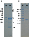
Fig. 1
Fig. 1
SDS-PAGE of the isolated cell envelope fraction. When characterized by SDS-PAGE, the cell envelope samples resolved in a typical pattern of five bands. The lane labelled with CE indicates the cell envelope samples, and the main bands are numbered from 1 to 5, with the bands 4 and 5 being the most represented; the asterisks indicate the running/stacking gel interface (*), the lysozyme band used for the peeling procedure (**), and the separation front with pigments (***), respectively. The lane labelled with M indicates the molecular marker. The sample shown here is the total cell envelope pool immediately after lysozyme digestion and before any further step; SDS-PAGE of the selectively released fraction. The selectively released fraction (SF) of the cell envelope pool was collected by centrifugation after lysozyme digestion and washed/concentrated with a 100 kDa cutoff concentrator. This procedure led to a complete removal of lysozyme. The selectively released fraction resolved in a single band (lane SF). The lane labelled with M indicates the molecular marker. The gels are cropped for clarity purpose
Fig. 2
Fig. 2
SEC profiles of the selectively released cell envelope fraction in absence and presence of DDM. In absence of DDM the protein samples appear characterized by two peaks a main one at high molecular weight (apparent mass > 800 kDa) and other secondary peaks at lower apparent molecular masses (continuous line). In presence of β-DDM the protein samples resolve into a single peak at an apparent mass of ~ 60 kDa followed by a shoulder (dashed line). The SDS-PAGE of the main peak for the SEC without DDM (-DDM) and with DDM (+ DDM) are also shown; the lane labelled with M indicates the molecular marker (Fig. 2, inset). The blue dots and the associated number represent the Log Molecular Weight (mass) of the molecular marker expressed in kDa. The gel is cropped for clarity purpose
Fig. 3
Fig. 3
Structure prediction and cryo-EM micrograph of the OMPb-like protein. The structure of the OMPb-like protein as predicted by Alpha-Fold. In (a), a top view with a detail of the pore is shown; in (b), a side view displaying a typical porin-like structure with a 8 stranded β-barrel organization is shown. The α-helix represents the signal peptide region. A typical grid field of the isolated protein visualized by cryo-EM (c). Several orientations of the particle are shown (yellow rings) including evident top views (yellow rings with dark dots). The particle is typically lengthened with one dimension dominating on the other two. The scale bar in (a) and (b) indicates 10 Å, while the scale bar in (c) indicates 100 Å
Fig. 4
Fig. 4
Characterization of the sample by UV-Vis absorption spectroscopyOMPb-like samples showed a typical and intense absorption in the UV range related with the protein backbone. In the visible region, it is evident the “three-fingers” trace related to the polyene-absorbing region typical for carotenoids. The inset shows a detail of the visible region with three bands at 463, 485, and 524 nm (black dashed line) and its comparison with the salinixanthin profile shown by Lanyi and Balashov (2008) (red dotted line)

Similar articles

References

    1. Allers T, Ngo HP. Genetic analysis of homologous recombination in Archaea: Haloferax volcanii as a model organism. Biochem Soc Trans. 2003;31:706–710. doi: 10.1042/bst0310706. - DOI - PubMed
    1. Antón J, Oren A, Benlloch S, Rodríguez-Valera F, Amann R, Rosselló-Mora R. Salinibacter ruber gen. nov., sp. nov., a novel, extremely halophilic member of the Bacteria from saltern crystallizer ponds. Int J Syst Evol Microbiol. 2002;52:485–491. doi: 10.1099/00207713-52-2-485. - DOI - PubMed
    1. Antón J, Peña A, Santos F, Martínez-García M, Schmitt-Kopplin P, Rosselló-Mora R. Distribution, abundance and diversity of the extremely halophilic bacterium Salinibacter ruber. Saline Syst. 2008;4:15. doi: 10.1186/1746-1448-4-15. - DOI - PMC - PubMed
    1. Balashov SP, Imasheva ES, Boichenko VA, Antón J, Wang JM, Lanyi JK. Xanthorhodopsin: a proton pump with a light-harvesting carotenoid antenna. Science. 2005;309:2061–2064. doi: 10.1126/science.1118046. - DOI - PMC - PubMed
    1. Balashov SP, Imasheva ES, Lanyi JK. Induced chirality of the light-harvesting carotenoid salinixanthin and its interaction with the retinal of xanthorhodopsin. Biochemistry. 2006;45:10998–11004. doi: 10.1021/bi061098i. - DOI - PMC - PubMed

Publication types

Supplementary concepts

LinkOut - more resources