Overexpression of mouse prion protein in transgenic mice causes a non-transmissible spongiform encephalopathy
- PMID: 36229637
- PMCID: PMC9562354
- DOI: 10.1038/s41598-022-21608-3
Overexpression of mouse prion protein in transgenic mice causes a non-transmissible spongiform encephalopathy
Abstract
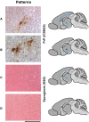
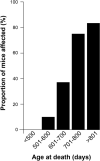
Transgenic mice over-expressing human PRNP or murine Prnp transgenes on a mouse prion protein knockout background have made key contributions to the understanding of human prion diseases and have provided the basis for many of the fundamental advances in prion biology, including the first report of synthetic mammalian prions. In this regard, the prion paradigm is increasingly guiding the exploration of seeded protein misfolding in the pathogenesis of other neurodegenerative diseases. Here we report that a well-established and widely used line of such mice (Tg20 or tga20), which overexpress wild-type mouse prion protein, exhibit spontaneous aggregation and accumulation of misfolded prion protein in a strongly age-dependent manner, which is accompanied by focal spongiosis and occasional neuronal loss. In some cases a clinical syndrome developed with phenotypic features that closely resemble those seen in prion disease. However, passage of brain homogenate from affected, aged mice failed to transmit this syndrome when inoculated intracerebrally into further recipient animals. We conclude that overexpression of the wild-type mouse prion protein can cause an age-dependent protein misfolding disorder or proteinopathy that is not associated with the production of an infectious agent but can produce a phenotype closely similar to authentic prion disease.
© 2022. The Author(s).
Conflict of interest statement
The authors declare that they have no conflicts of interest relevant to this manuscript, with the exceptions of: J.C. is a Director and G.S.J., J.D.F.W. and J.C. are shareholders of D-Gen Limited (London), an academic spin-out company working in the field of prion disease diagnosis, decontamination and therapeutics. D-Gen supplied the antibody ICSM35 used in this study.
Figures


Similar articles
-
PrP aggregation can be seeded by pre-formed recombinant PrP amyloid fibrils without the replication of infectious prions.Acta Neuropathol. 2016 Oct;132(4):611-24. doi: 10.1007/s00401-016-1594-5. Epub 2016 Jul 4. Acta Neuropathol. 2016. PMID: 27376534 Free PMC article.
-
Recombinant Mammalian Prions: The "Correctly" Misfolded Prion Protein Conformers.Viruses. 2022 Aug 31;14(9):1940. doi: 10.3390/v14091940. Viruses. 2022. PMID: 36146746 Free PMC article. Review.
-
Dissociation of prion protein amyloid seeding from transmission of a spongiform encephalopathy.J Virol. 2013 Nov;87(22):12349-56. doi: 10.1128/JVI.00673-13. Epub 2013 Sep 11. J Virol. 2013. PMID: 24027305 Free PMC article.
-
Inherited prion disease A117V is not simply a proteinopathy but produces prions transmissible to transgenic mice expressing homologous prion protein.PLoS Pathog. 2013;9(9):e1003643. doi: 10.1371/journal.ppat.1003643. Epub 2013 Sep 26. PLoS Pathog. 2013. PMID: 24086135 Free PMC article.
-
The use of transgenic mice in the investigation of transmissible spongiform encephalopathies.Rev Sci Tech. 1998 Apr;17(1):278-90. doi: 10.20506/rst.17.1.1079. Rev Sci Tech. 1998. PMID: 9638817 Review.
Cited by
-
Convergent generation of atypical prions in knockin mouse models of genetic prion disease.J Clin Invest. 2024 Aug 1;134(15):e176344. doi: 10.1172/JCI176344. J Clin Invest. 2024. PMID: 39087478 Free PMC article.
-
Development of a sensitive real-time quaking-induced conversion (RT-QuIC) assay for application in prion-infected blood.PLoS One. 2023 Nov 2;18(11):e0293845. doi: 10.1371/journal.pone.0293845. eCollection 2023. PLoS One. 2023. PMID: 37917783 Free PMC article.
References
Publication types
MeSH terms
Substances
Grants and funding
LinkOut - more resources
Full Text Sources
Medical
Molecular Biology Databases

