

Comparison of in vivo hindfoot joints motion changes during stance phase between non-flatfoot and stage II adult acquired flatfoot
- PMID: 36229819
- PMCID: PMC9559000
- DOI: 10.1186/s13047-022-00577-w
Comparison of in vivo hindfoot joints motion changes during stance phase between non-flatfoot and stage II adult acquired flatfoot
Abstract
Background: To compare the kinematic characteristics of hindfoot joints in stage II adult acquired flatfoot deformity (AAFD) with those of non-flatfoot through the 3D-to-2D registration technology and single fluoroscopic imaging system.
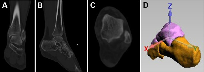
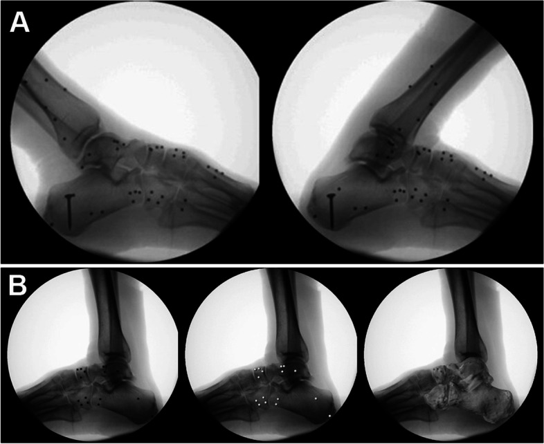
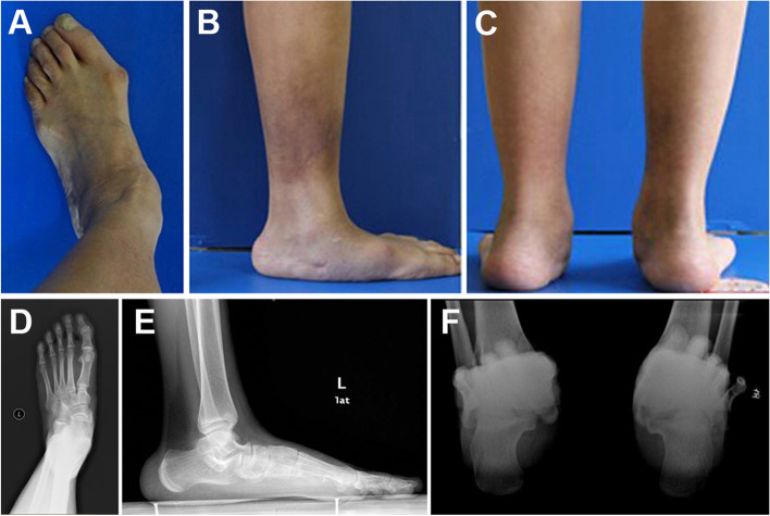
Methods: Eight volunteers with stage II AAFD and seven volunteers without stage II AAFD were recruited and CT scans were performed bilateral for both groups in neutral positions. Their lateral dynamic X-ray data during the stance phase, including 14 non-flatfeet and 10 flatfeet, was collected. A computer-aided simulated light source for 3D CT model was applied to obtain the virtual images, which were matched with the dynamic X-ray images to register in the "Fluo" software, so that the spatial changes during the stance phase could be calculated.
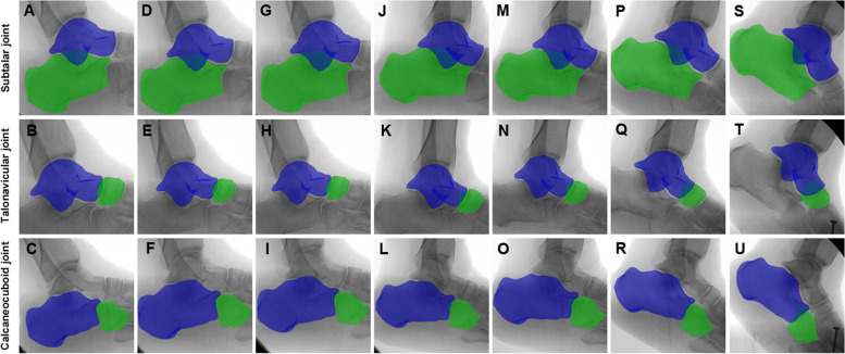
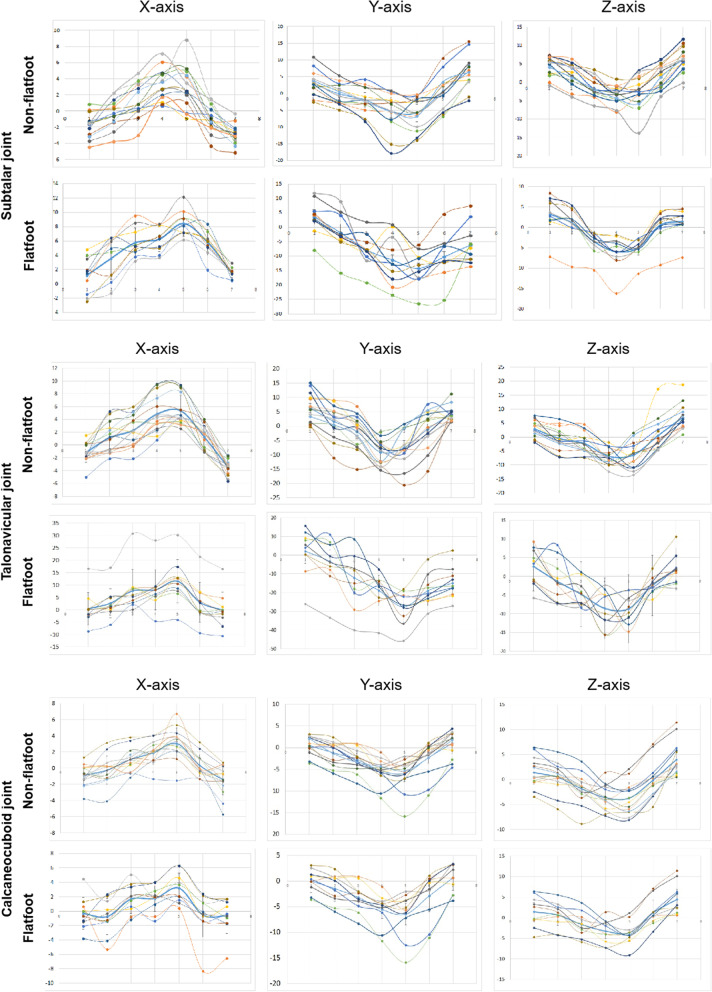
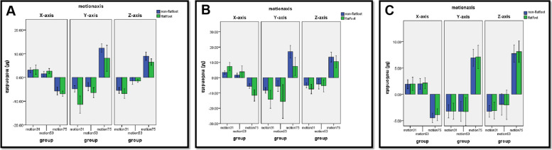
Results: During the early-stance phase, the calcaneous was more dorsiflexed, everted, and externally-rotated relative to the talus in flatfoot compared with that in non-flatfoot (p < 0.05). During the mid-stance phase, the calcaneous was more dorsiflexed and everted relative to the talus in flatfoot compared with that in non-flatfoot (p < 0.05); however, the rotation did not differ significantly between the two groups (p > 0.05). During the late-stance phase, the calcaneous was more plantarflexed, but less inverted and internally-rotated, relative to the talus in flatfoot compared with that in non-flatfoot (p < 0.05). During the early- and mid-stance phase, the navicular was more dorsiflexed, everted, and externally-rotated relative to the talus in flatfoot compared with that in non-flatfoot (p < 0.05). During the late-stance phase, the navicular was more plantarflexed, but less inverted and internally-rotated, relative to the talus in flatfoot compared with that in non-flatfoot (p < 0.05). There was no difference in the motion of cuboid between the two groups during the whole stance phase (p > 0.05).
Conclusions: During the early- and mid-stance phase, excessive motion was observed in the subtalar and talonavicular joints in stage II AAFD. During the late-stance phase, the motion of subtalar and talonavicular joints appeared to be in the dysfunction state. The current study helps better understanding the biomechanics of the hindfoot during non-flatfoot and flatfoot condition which is critical to the intervention to the AAFD using conservative treatment such as insole or surgical treatment for joint hypermotion.
Keywords: Adult acquired flatfoot deformity; Hindfoot joint; In vivo; Movement.
© 2022. The Author(s).
Conflict of interest statement
The authors declare that they have no competing interests.
Figures

Similar articles
-
Pathological kinematic patterns of the tarsal complex in stage II adult-acquired flatfoot deformity.J Orthop Res. 2019 Feb;37(2):477-482. doi: 10.1002/jor.23821. Epub 2019 Feb 1. J Orthop Res. 2019. PMID: 29194779
-
An in vivo study of hindfoot 3D kinetics in stage II posterior tibial tendon dysfunction (PTTD) flatfoot based on weight-bearing CT scan.Bone Joint Res. 2013 Dec 9;2(12):255-63. doi: 10.1302/2046-3758.212.2000220. Print 2013. Bone Joint Res. 2013. PMID: 24324193 Free PMC article.
-
Three-dimensional kinematic change of hindfoot during full weightbearing in standing: an analysis using upright computed tomography and 3D-3D surface registration.J Orthop Surg Res. 2019 Nov 11;14(1):355. doi: 10.1186/s13018-019-1443-z. J Orthop Surg Res. 2019. PMID: 31711523 Free PMC article.
-
Talus morphology differs between flatfeet and controls, but its variety has no influence on extent of surgical deformity correction.Arch Orthop Trauma Surg. 2022 Nov;142(11):3103-3110. doi: 10.1007/s00402-021-03925-w. Epub 2021 May 10. Arch Orthop Trauma Surg. 2022. PMID: 33970321 Free PMC article. Review.
-
Operative treatment of the difficult stage 2 adult acquired flatfoot deformity.Foot Ankle Clin. 2001 Mar;6(1):95-119. doi: 10.1016/s1083-7515(03)00083-4. Foot Ankle Clin. 2001. PMID: 11385931 Review.
References
-
- Raduan FC, Coetzee JC, Den Hartog BD, Seybold JD, Cammack PM, Stone RM, et al. A new approach for stage 2 adult acquired flatfoot deformity. Foot Ankle Orthop. 2022;7(1):2473011421S00406. doi: 10.1177/2473011421S00406. - DOI
MeSH terms
Grants and funding
- 81902303/National Natural Science Foundation of China
- 82102632/National Natural Science Foundation of China
- 82160412/National Natural Science Foundation of China
- 2020A151501048/Guangdong Basic and Applied Basic Research Foundation
- B2019085/Guangdong Medical Research Foundation
- 2020GXNSFBA297089/Guangxi Natural Science Foundation
- JCYJ20190806164216661/Shenzhen Science and Technology Project
- RCBS20200714114856299/Shenzhen Science and Technology Project
- 2021CBB0106/Liuzhou Science and Technology Project
- 20203357028/Clinical Research Project of Shenzhen Second People's Hospital
LinkOut - more resources
Full Text Sources
