. 2022 Oct 13;15(1):365.
doi: 10.1186/s13071-022-05500-7.

Molecular insights into the heat shock proteins of the human parasitic blood fluke Schistosoma mansoni

Affiliations

Molecular insights into the heat shock proteins of the human parasitic blood fluke Schistosoma mansoni

Nancy A Aguoru et al. Parasit Vectors. .

Abstract

Background: Heat shock proteins (HSPs) are evolutionarily conserved proteins, produced by cells in response to hostile environmental conditions, that are vital to organism homeostasis. Here, we undertook the first detailed molecular bioinformatic analysis of these important proteins and mapped their tissue expression in the human parasitic blood fluke, Schistosoma mansoni, one of the causative agents of the neglected tropical disease human schistosomiasis.

Methods: Using bioinformatic tools we classified and phylogenetically analysed HSP family members in schistosomes, and performed transcriptomic, phosphoproteomic, and interactomic analysis of the S. mansoni HSPs. In addition, S. mansoni HSP protein expression was mapped in intact parasites using immunofluorescence.

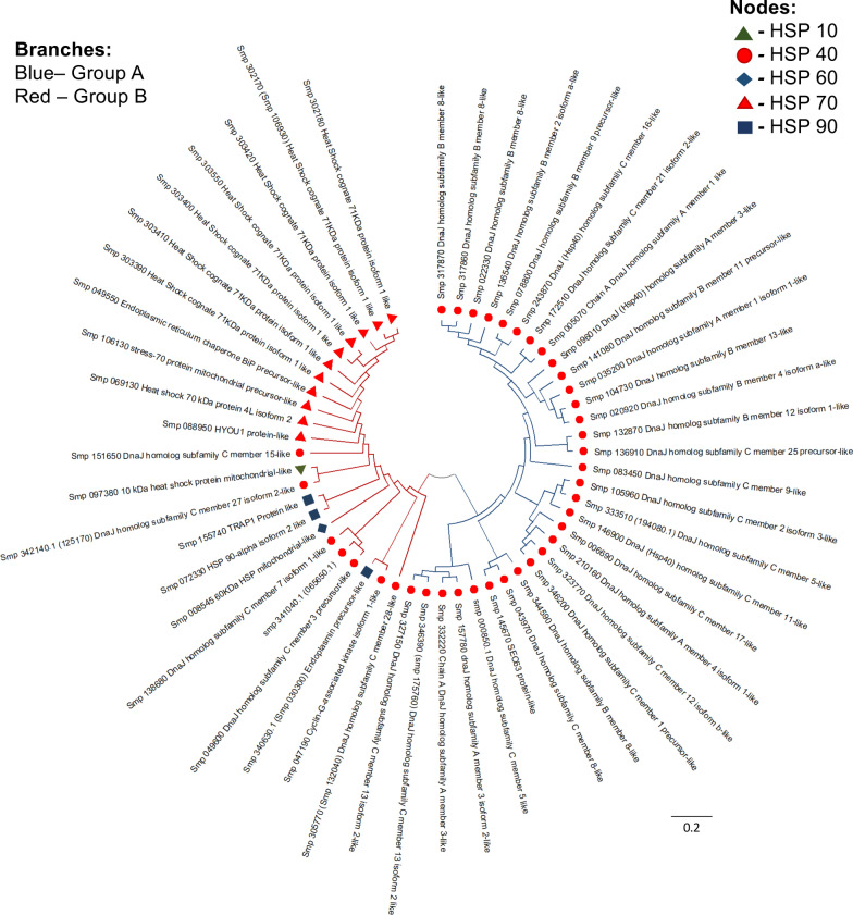
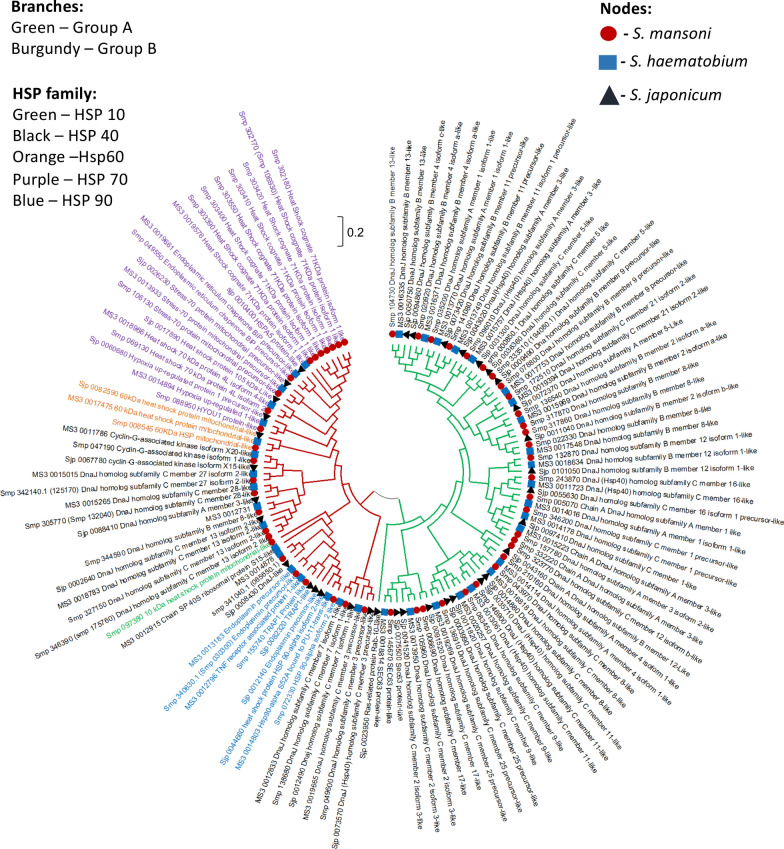
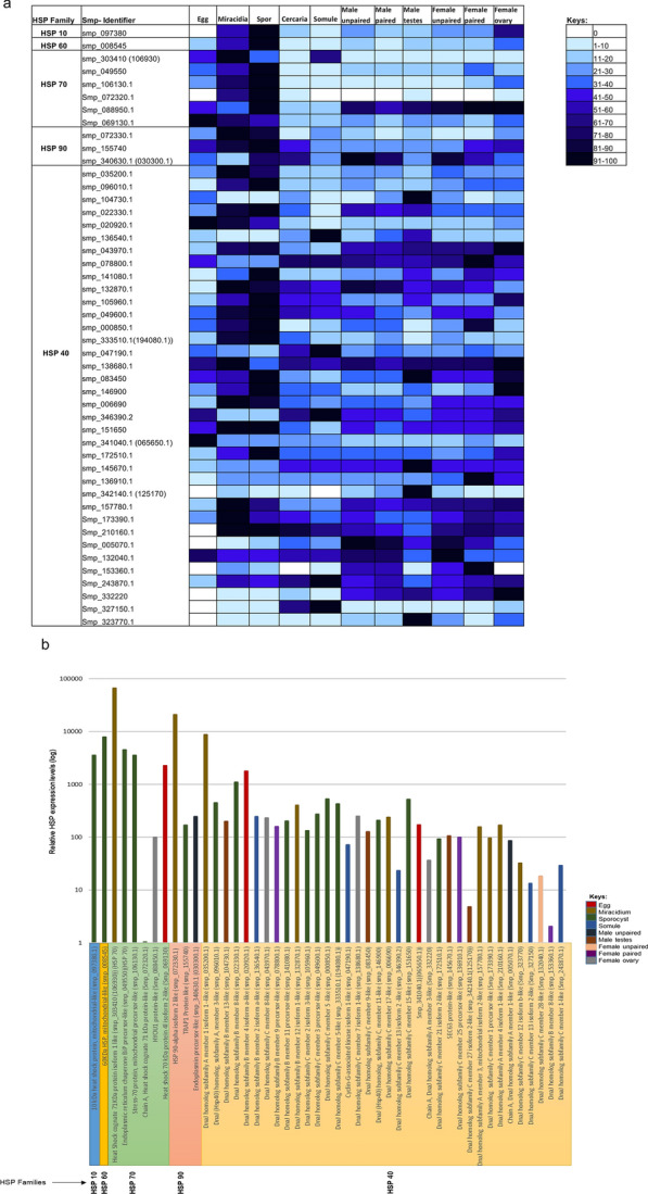
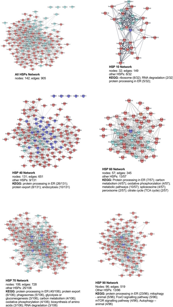
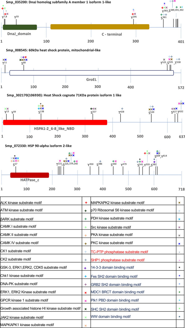
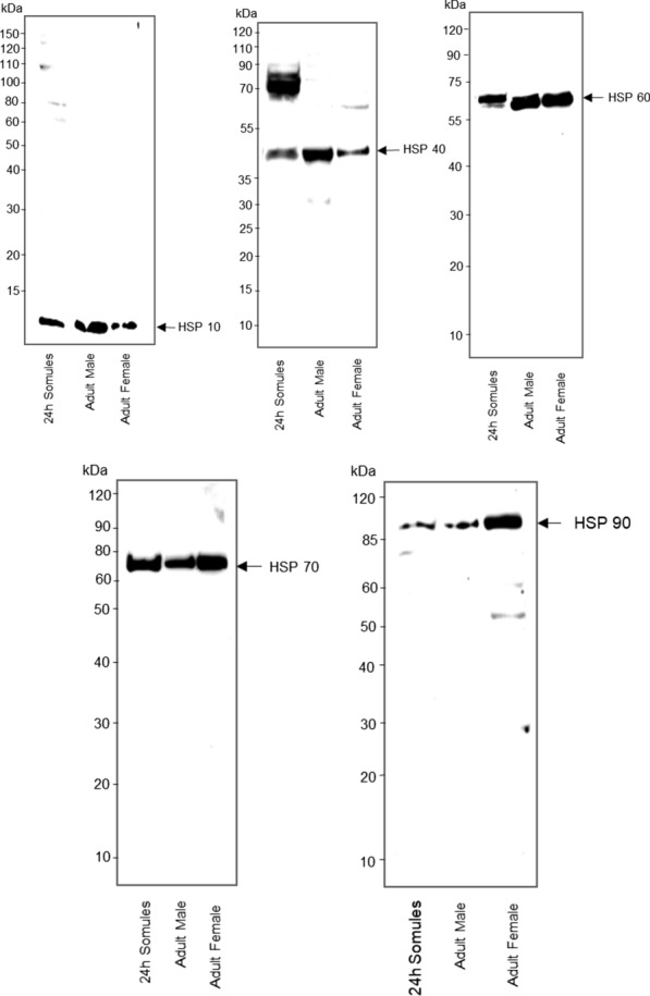
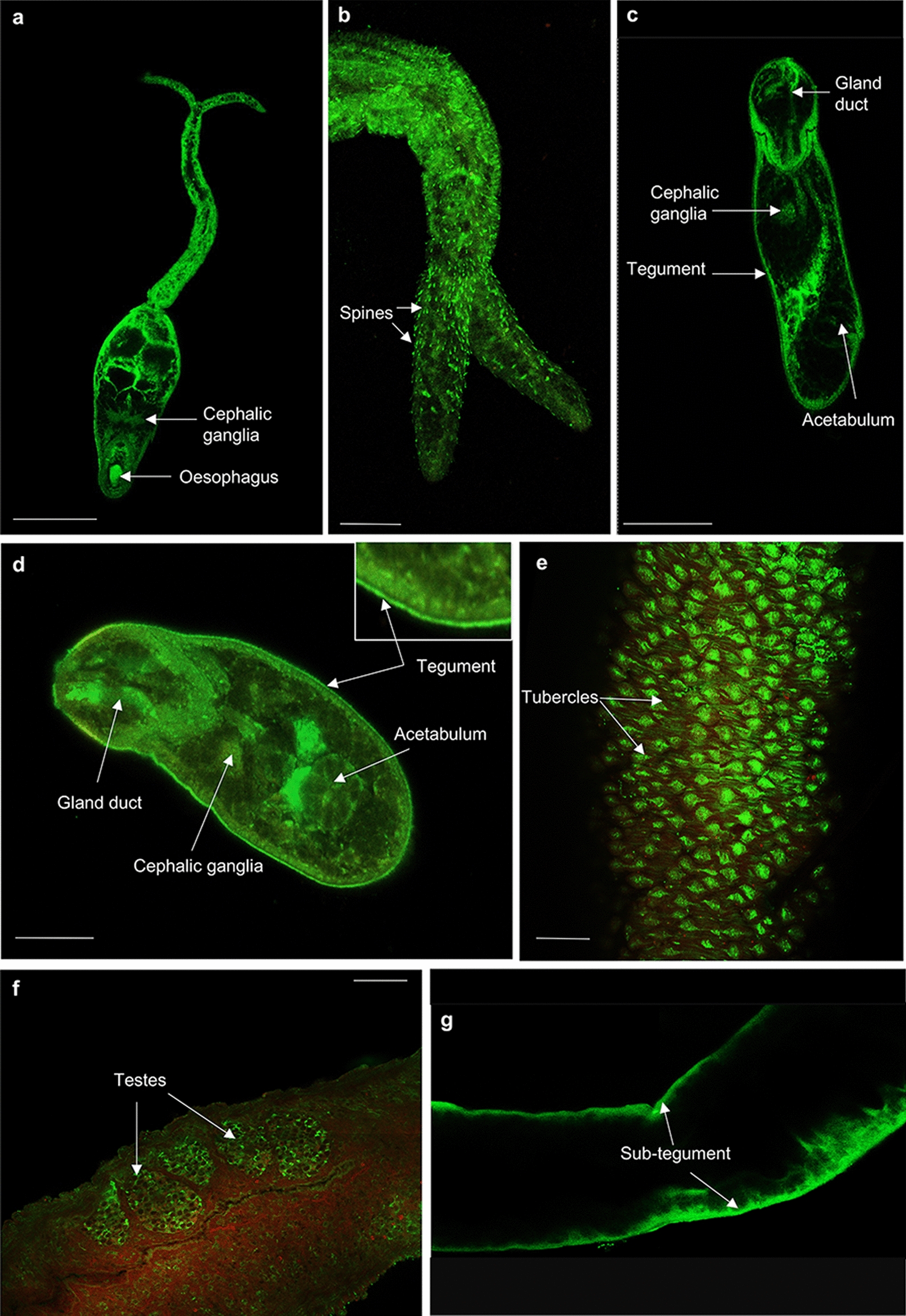
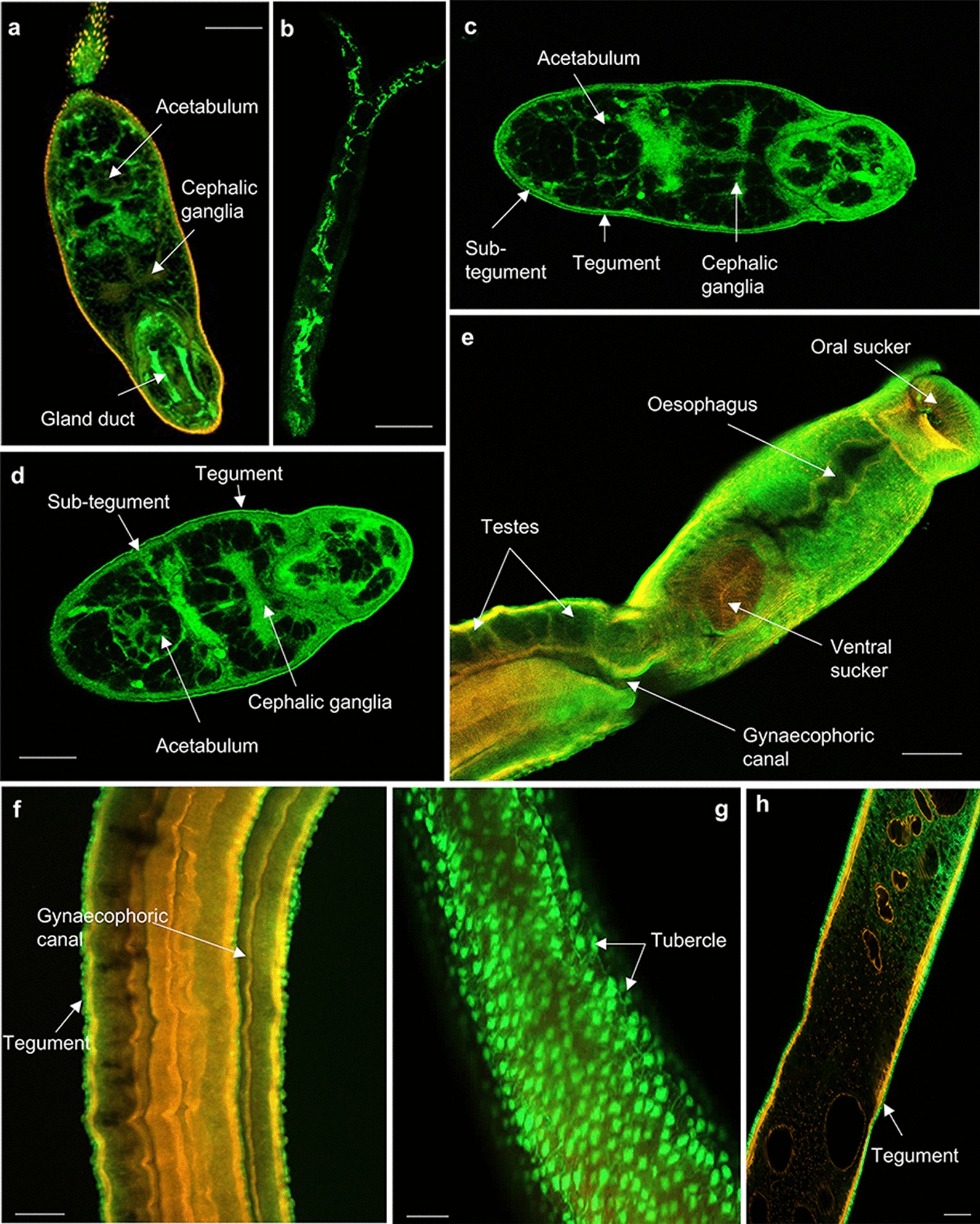
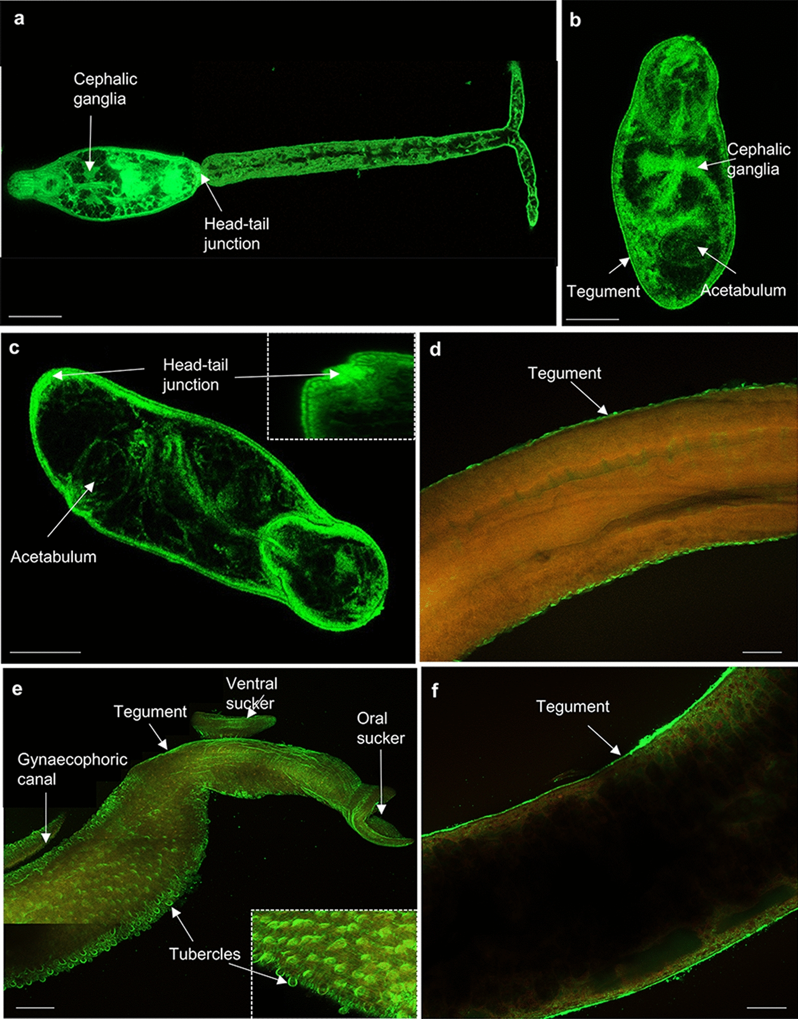
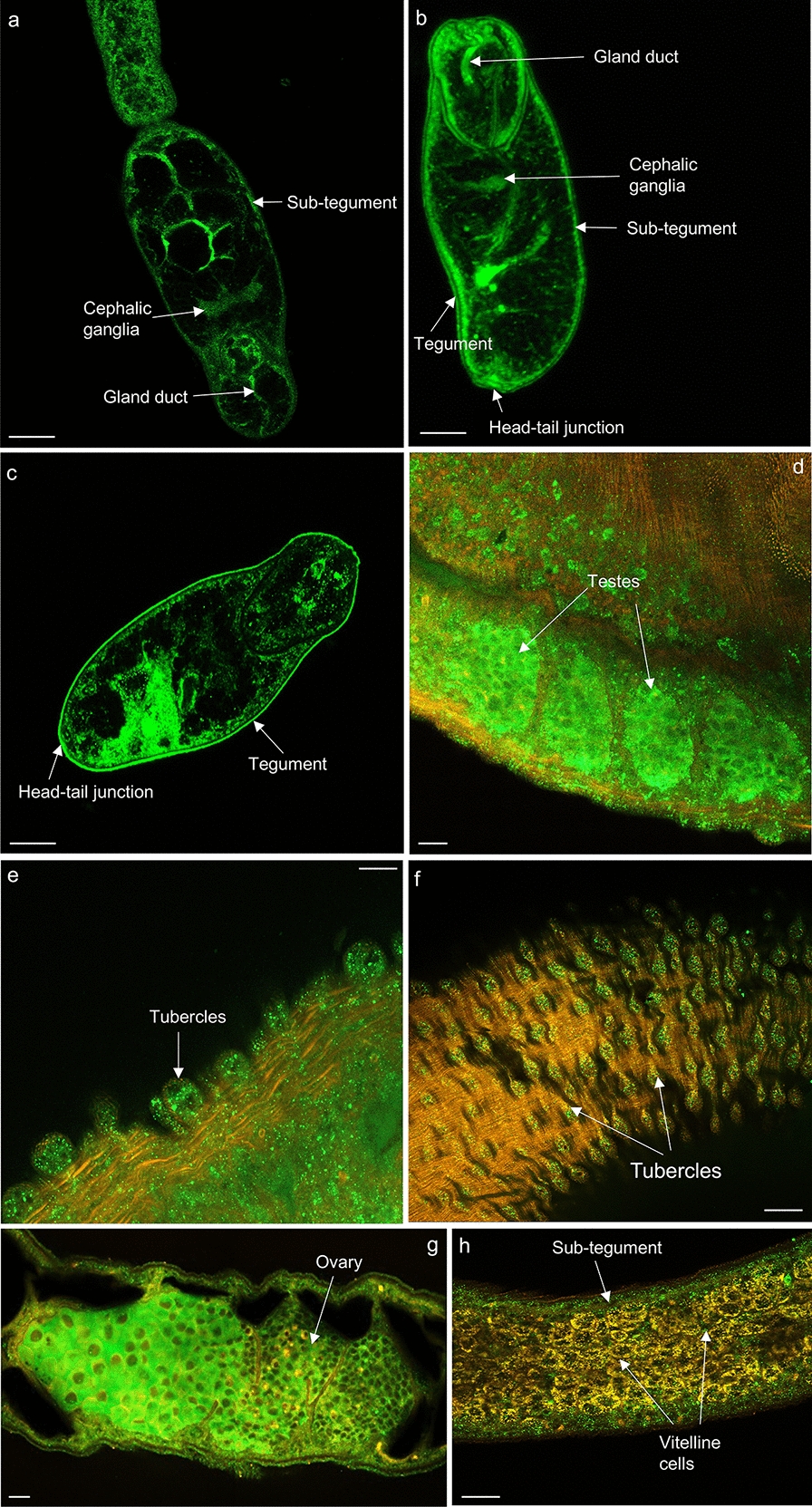
Results: Fifty-five HSPs were identified in S. mansoni across five HSP families; high conservation of HSP sequences were apparent across S. mansoni, Schistosoma haematobium and Schistosoma japonicum, with S. haematobium HSPs showing greater similarity to S. mansoni than those of S. japonicum. For S. mansoni, differential HSP gene expression was evident across the various parasite life stages, supporting varying roles for the HSPs in the different stages, and suggesting that they might confer some degree of protection during life stage transitions. Protein expression patterns of HSPs were visualised in intact S. mansoni cercariae, 3 h and 24 h somules, and adult male and female worms, revealing HSPs in the tegument, cephalic ganglia, tubercles, testes, ovaries as well as other important organs. Analysis of putative HSP protein-protein associations highlighted proteins that are involved in transcription, modification, stability, and ubiquitination; functional enrichment analysis revealed functions for HSP networks in S. mansoni including protein export for HSP 40/70, and FOXO/mTOR signalling for HSP90 networks. Finally, a total of 76 phosphorylation sites were discovered within 17 of the 55 HSPs, with 30 phosphorylation sites being conserved with those of human HSPs, highlighting their likely core functional significance.

Conclusions: This analysis highlights the fascinating biology of S. mansoni HSPs and their likely importance to schistosome function, offering a valuable and novel framework for future physiological investigations into the roles of HSPs in schistosomes, particularly in the context of survival in the host and with the aim of developing novel anti-schistosome therapeutics.

Keywords: Gene expression; Heat shock protein 10; Heat shock protein 40; Heat shock protein 70; Heat shock protein 90; Heat shock proteins; Phosphorylation; Protein expression; Protein–protein association network; Schistosome.


Conflict of interest statement

AJW is a member of the editorial board of Parasites and Vectors. The other authors declare that they have no competing interests.

Figures

Fig. 1
Fig. 1
Comparative analysis of the total number of heat shock proteins (HSPs) identified in humans, free-living flatworms and Schistosoma species. HSPs were identified through bioinformatic similarity searches for each HSP family (HSP 10, HSP 40, HSP 60, HSP 70 and HSP 90)
Fig. 2
Fig. 2
Phylogenetic tree revealing the evolutionary history of Schistosoma mansoni HSPs. The evolutionary history of the HSP 10, 40, 60, 70 and 90 families was inferred by neighbour-joining method analysis [74]. Evolutionary distances were computed using the Poisson correction method [75]. The analysis involved 54 amino acid sequences (one each of HSP 10 and 60, eleven of HSP 70, three of HSP 90 and 38 of HSP 40); one HSP 70 member was excluded as it contained significant gaps in the alignment. The analysis (unrooted circular tree) was conducted using Molecular Evolutionary Genetic Analysis (MEGA) X [76]
Fig. 3
Fig. 3
Phylogenetic tree of the HSPs from Schistosoma mansoni, Schistosoma haematobium and Schistosoma japonicum. The evolutionary history was inferred by neighbour-joining method analysis [74] and the distance computed using the Poisson correction method [75]. The analysis involved 127 amino acid sequences. The evolutionary relationships (unrooted circular tree) were determined using MEGA X [76]
Fig. 4
Fig. 4
a, b HSP gene expression in Schistosoma mansoni during development. a Comparative analysis of the expression profiles of different HSPs during development of the parasite from egg to adult worm using quantitative data obtained from Schisto.xyz. Ten percentile expression categories were calculated from the gene expression values relative to the maximum expression level for each gene that was assigned 100%. White represents no expression and black represents maximum (100%) expression for each gene. b Comparative analysis of the maximum HSP gene expression data and the associated parasite life stage for each HSP member; note that “cercaria” and “male paired” are absent as no HSP gene was maximally expressed at these stages
Fig. 5
Fig. 5
Schistosoma mansoni HSP interaction networks. Identified S. mansoni HSPs were mapped using the Search Tool for the Retrieval of Interacting Genes/Proteins (STRING) database of predicted protein–protein interactions, embedded in Cytoscape for all HSPs and specific HSP (HSP 10, HSP 40, HSP 60, HSP 70 and HSP 90) families using a maximum of 100 additional interactors (proteins, nodes) for each query (seed) protein. Mapping of interactions (edges) was performed at high (0.7) confidence. Proteins coloured red within each network represent non-HSPs, with light blue and dark blue representing “other” HSPs and the HSP seed node, respectively. Out of a total of 55 S. mansoni HSPs, 42 were identified by the STRING database and were included in the analysis. The number of proteins identified per network that feature in a specific Kyoto Encyclopedia of Genes and Genomes (KEGG) pathway is indicated for each network. ER Endoplasmic reticulum
Fig. 6
Fig. 6
Phosphorylation sites within Schistosoma mansoni HSPs. Data on the phosphorylation sites of S. mansoni HSPs were mined from the S. mansoni phosphoproteome published by Hirst et al. [54]. Four HSPs are shown; the remainder can be viewed in Additional file 4: Figure S1. Domains within each HSP were identified using the conserved protein domain tool within InterPro and the National Center for Biotechnology Information (NCBI) Conserved Domains Database. Putative upstream kinases, phosphatases and binding motifs were identified using the Human Protein Reference Database (HPRD) motif finder and the Phosphorylation Site Database (PHOSIDA). Kinases are written in black and are denoted by a plus or a cross symbol, phosphatases are in red and are denoted by a minus symbol, binding motifs are in blue and are denoted by an asterisk
Fig. 7
Fig. 7
Detection of HSPs in Schistosoma mansoni. In vitro 24-h-cultured somules (~ 1000 per lane) or adult worms (one male or female per lane) were homogenised and protein extracts subjected to western blotting with one of the following antibodies: anti-HSPE1 (ARPP54651_P050) (HSP 10) raised against a synthetic peptide located within amino acid 55–102 of human HSPE1; anti-HSP 40 (SPC-100), which recognises the whole amino acid sequence of human DNAJB1; anti-HSP 60 (SMC-111A/B) raised against amino acid sequence 382–447 of human HSP 60; anti-HSP 70 (SMC-162 C/D) raised against the whole amino acid sequence of human heat shock 70 kDa protein 1A and heat shock 70 kDa protein 1B; or anti-HSP 90 (STJ93608) that recognises amino acids 180–260 of human HSP 90β
Fig. 8
Fig. 8
ag Mapping HSP 10 in Schistosoma mansoni. Cercariae, somules, male and female adult worms were fixed and processed for immunofluorescence using anti-HSPE1 (HSP 10) primary and Alexa Fluor 488 secondary (green) antibodies; rhodamine phalloidin (red) was used to stain filamentous actin. Samples were mounted on slides and images captured on a Zeiss LSM 800 confocal laser scanning microscope. HSP 10 localised in a cercariae; b 3-h somules; c 24-h somules; d, e adult males; and f, g adult females. Representative micrographs are single z-sections through the parasite. Scale bars = 25 µm (for cercariae and somules) and 50 µm (for adult worms)
Fig. 9
Fig. 9
ag Mapping HSP 60 in Schistosoma mansoni. Cercariae, somules, male and female adult worms were fixed and processed for immunofluorescence using anti-HSP 60 primary and Alexa Fluor 488 secondary antibody (green) antibodies; rhodamine phalloidin (red) was used to stain filamentous actin. Samples were mounted on slides and images captured on a Zeiss LSM 800 confocal microscope. HSP 60 localised in a, b cercariae; c 3-h somules; d 24-h somules; e, f adult males; and g adult females. Representative micrographs are single z-sections through the parasite. Scale bars = 25 µm (for cercariae and somules) and 50 µm (for adult worms)
Fig. 10
Fig. 10
ah Mapping HSP 40 in Schistosoma mansoni. Cercariae, somules, male and female adult worms were fixed and processed for immunofluorescence using anti-HSP 40 primary and Alexa Fluor 488 secondary antibodies (green); rhodamine phalloidin (red) was used to stain filamentous actin. Samples were mounted on slides and images captured on a Zeiss LSM 800 confocal microscope. HSP 40 localised in a, b cercariae; c 3-h somules; d 24-h somules; eg adult males; and h adult females. Representative micrographs are single z-sections through the parasite. Scale bars = 25 µm (for cercariae and somules) and 50 µm (for adult worms)
Fig. 11
Fig. 11
ag Mapping HSP 70 in Schistosoma mansoni. Cercariae, somules, and male and female adult worms were fixed and processed for immunofluorescence using anti-HSP 70 primary and Alexa Fluor 488 secondary antibodies (green); rhodamine phalloidin (red) was used for filamentous actin staining. Samples were mounted on slides and images captured using a Zeiss LSM 800 confocal microscope. HSP 70 localised in a cercariae; b 3-h somules; c 24-h somules; d, e adult males; and f adult females. Representative images are eg maximum projections of confocal z-stacks; the rest are single z-sections through the parasite. Scale bars = 25 µm (for cercariae and somules) and 50 µm (for adult worms)
Fig. 12
Fig. 12
ah Mapping of HSP 90 in Schistosoma mansoni. Cercariae, somules, male and female adult worms were fixed and processed for immunohistochemistry using anti-HSP 90 primary and Alexa Fluor 488 secondary antibodies (green); rhodamine phalloidin (red) was used for filamentous actin staining. Samples were mounted on slides and images captured on a Zeiss LSM 800 confocal microscope. HSP 90 localised in the a cercariae; b 3-h somules; c 24-h somules; df adult males; and g, h adult females. Representative micrographs are single z-sections through the parasite. Scale bars = 25 µm (for cercariae and somules) and 50 µm (for adult worms)

Similar articles

Cited by

References

    1. Berriman M, Haas BJ, Loverde PT, Wilson RA, Dillon GP, Cerqueira GC, et al. The genome of the blood fluke Schistosoma mansoni. Nature. 2009;460:352–358. - PMC - PubMed
    1. Walker A. Insights into the functional biology of schistosomes. Parasite Vectors. 2011;4:203. - PMC - PubMed
    1. World Health Organization . Schistosomiasis: control report 2001–2011 and strategic plan 2012–2020. Geneva: WHO Press; 2013.
    1. Engels D, Chitsulo L, Montresor A, Savioli L. The global epidemiological situation of schistosomiasis and new approaches to control and research. Acta Trop. 2002;82:139–146. - PMC - PubMed
    1. Gray DJ, Ross AG, Li YS, McManus DP. Diagnosis and management of schistosomiasis. Br Med J. 2011;342:d2651. - PMC - PubMed

Substances

LinkOut - more resources