. 2022 Sep 21;12(19):2508.
doi: 10.3390/ani12192508.

RNA Sequencing Reveals the Regulation of Betaine on Chicken Myogenesis

Affiliations

RNA Sequencing Reveals the Regulation of Betaine on Chicken Myogenesis

Zhijun Wang et al. Animals (Basel). .

Abstract

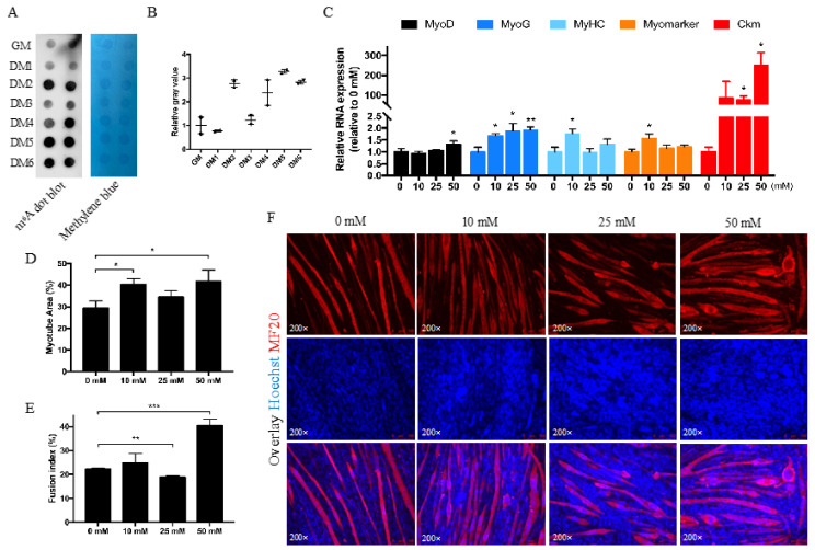
Betaine is trimethylglycine and a universal methyl donor which could provide methyl and glycine for cells and animals. As a new star in epigenetics, N6-Methyladenosine has been reported to regulate multiple biological activities, but the regulatory mechanism of betaine on N6-Methyladenosine as well as myogenesis was little studied. In this study, we treated chicken primary myoblast cells with different concentrations of betaine (0, 10, 25, and 50 mmol/L) and found that myoblast cell proliferation was inhibited, although the cell cycle was promoted in the S phase by betaine, where the myotube area was increased as well as the differentiation marker genes MyoD, MyoG, MyHC, Myomarker, and Ckm. RNA sequencing obtained a total of 61 differentially expressed genes (DEGs); DEGs caused by 50 mmol/L betaine were mainly enriched in the regulation of skeletal muscle tissue regeneration and some amino acid metabolic processes. The gene expression pattern trends of all DEGs were mainly clustered into 2 profiles, with the increase in betaine concentration, the gene expression pattern either increased or decreased continuously. Overall, a low concentration betaine can increase the N6-Methyladenosine modification level and myotube area but depresses myoblast cell proliferation in vitro.

Keywords: N6-Methyladenosine; betaine; chicken; mRNA-Seq; myogenesis.


Conflict of interest statement

The authors declare no conflict of interest.

Figures

Figure 1
Figure 1
Betaine inhibits myoblast cells proliferation. (A) RNA dot blot and methylene blue results after being treated with 0, 10, 25, 50 mmol/L betaine for 24 h. (B) The relative gray value of RNA dot blot. (C) Cell cycle analysis of different cell stages after 24 h betaine treatment. (D) The statistical results of CCK-8 assay after 24 h, 36 h, and 48 h betaine treatment. (E) The EdU positive cell rate (F) and EdU staining of myoblast cells being treated with betaine. (mM refers to mmol/L; * p < 0.05; ** p < 0.01; *** p < 0.001).
Figure 2
Figure 2
Betaine promotes myotube formation. (A) RNA dot blot and methylene blue results during myoblast differentiation. (B) The relative gray value of RNA dot blot. (C) qRT-PCR results for MyoD, MyoG, MyHC, Myomarker, and Ckm in myoblast cells after being treated with 0, 10, 25, 50 mmol/L betaine for 24 h. (DF) Myotube area (%) analysis (D), fusion index € of MF20 immunofluorescence staining (F) after being treated with 0, 10, 25, 50 mmol/L betaine for 24 h at DM3. (mM refers to mmol/L; * p < 0.05; ** p < 0.01; *** p < 0.001).
Figure 3
Figure 3
Overview of mRNA sequencing in betaine-treated myoblast cells. (A) Venn diagram shows common and unique DEGs between groups. (B) The number of differentially up- and down-expressed genes in each group. (C) GO functional enrichment analysis of DEGs in Bet_0 vs. Bet_50. (D) KEGG pathway analysis of DEGs in Bet_0 vs. Bet_50. (E) Heatmap for all 61 DEGs.
Figure 4
Figure 4
Gene expression pattern analysis of all DEGs. (A) General chart of all gene expression trend profiles. (B) Gene expression trend profiles are ordered based on the p-value significance of the number of genes assigned versus expected. (C, D) the FPKM value (C) and qRT-PCR validation (D) of eight DEGs in profile 0 and 19. (mM refers to mmol/L; * p < 0.05; ** p < 0.01; *** p < 0.001.)

References

    1. Mitchell A.D., Chappell A., Knox K.L. Metabolism of betaine in the ruminant. J. Anim. Sci. 1979;49:764–774. doi: 10.2527/jas1979.493764x. - DOI - PubMed
    1. Lawrence B.V., Schinckel A.P., Adeola O., Cera K. Impact of betaine on pig finishing performance and carcass composition. J. Anim. Sci. 2002;80:475–482. doi: 10.2527/2002.802475x. - DOI - PubMed
    1. Zhao G., He F., Wu C., Li P., Li N., Deng J., Zhu G., Ren W., Peng Y. Betaine in Inflammation: Mechanistic Aspects and Applications. Front. Immunol. 2018;9:1070. doi: 10.3389/fimmu.2018.01070. - DOI - PMC - PubMed
    1. Kumar S., Cheng X., Klimasauskas S., Mi S., Posfai J., Roberts R.J., Wilson G.G. The DNA (cytosine-5) methyltransferases. Nucleic Acids Res. 1994;22:1–10. doi: 10.1093/nar/22.1.1. - DOI - PMC - PubMed
    1. Hori H. Methylated nucleosides in tRNA and tRNA methyltransferases. Front. Genet. 2014;5:144. doi: 10.3389/fgene.2014.00144. - DOI - PMC - PubMed