

Effect of Supplementation of Cryoprotectant Solution with Hydroxypropyl Cellulose for Vitrification of Bovine Oocytes
- PMID: 36230376
- PMCID: PMC9559640
- DOI: 10.3390/ani12192636
Effect of Supplementation of Cryoprotectant Solution with Hydroxypropyl Cellulose for Vitrification of Bovine Oocytes
Abstract
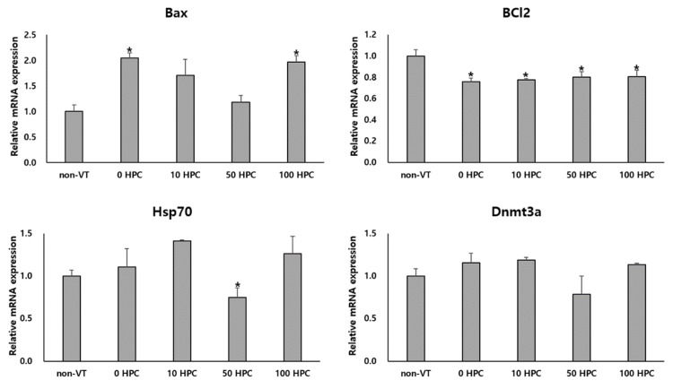
The technology of successful cryopreservation is a very important factor in research and commercial applications. However, the survival and development of the vitrified-thawed (VT) oocytes are lower than those of non-vitrified-thawed (non-VT) oocytes. This study investigated the effect of the addition of hydroxypropyl cellulose (HPC) to a vitrification solution of bovine oocytes. For the vitrification, bovine metaphase II oocytes were pretreated with a solution containing 10% ethylene glycol supplemented with 0, 10, 50, or 100 µg/mL HPC for 5 min, then exposed to a solution containing 30% ethylene glycol supplemented with 0, 10, 50, or 100 µg/mL HPC for 30 sec, and then directly plunged into liquid nitrogen. Oocytes exposed to 0, 10, 50, and 100 µg/mL HPC were named the 0, 10, 50, and 100 HPC groups, respectively. Samples were thawed via sequential incubation in Dulbecco's phosphate-buffered saline (D-BPS) supplemented with 10% fetal bovine serum and decreasing concentrations of sucrose (1, 0.5, 0.25, and 0.125 M) for 1 min each time. After thawing, VT oocytes were treated at 0.05% hyaluronidase, and cumulus cells were removed by mechanical pipetting. The oocytes were washed with HEPES-buffered Tyrode's medium and incubated in a droplet of previously cultured in vitro maturation medium for 1 h to recover. The survival rate of the oocytes was significantly higher in the 50 HPC group (84.2%) than in the 0 (75.4%), 10 (80.4%), and 100 (75.5%) HPC groups. The reactive oxygen species (ROS) levels of the non-VT and 50 HPC groups were lower than the 0, 10, and 100 HPC groups. The mRNA levels of proapoptotic genes (Bax) were lower in the non-VT, 0, and 50 HPC groups than in the other groups. The mRNA expression levels of antiapoptotic genes (BCl2) was higher in the non-VT than in the other groups. The mRNA level of a stress-related gene (Hsp70) was lower in the 50 HPC than in the other groups. At day 8, the developmental capacity of embryos obtained via parthenogenetic activation (PA) was determined in the non-VT, 0 HPC, and 50 HPC groups. The cleavage rate of the non-VT group was significantly higher, but the blastocyst development rate and total cell number per blastocyst did not significantly differ between the non-VT and 50 HPC groups. The mRNA levels of proapoptotic genes (Bax and Caspase-3) and a stress-related gene (Hsp70) were higher in the 0 HPC group than in the non-VT and 50 HPC groups. In conclusion, supplementation of vitrification solution with HPC improves the survival rate of VT bovine oocytes and the development capacity of embryos derived from these oocytes via PA.
Keywords: bovine oocytes; cryoprotectant; hydroxypropyl cellulose; solution vitrification.
Conflict of interest statement
The authors declare no conflict of interest.
Figures


References
-
- Sansinena M., Santos M.V., Chirife J., Zaritzky N. In-vitro development of vitrified-warmed bovine oocytes after activation may be predicted based on mathematical modelling of cooling and warming rates during vitrification, storage and sample removal. Reprod. Biomed. Online. 2018;36:500–507. doi: 10.1016/j.rbmo.2018.01.003. - DOI - PubMed
-
- Somfai T., Kikuchi K. Vitrification of porcine oocytes and zygotes in microdrops on a solid metal surface or liquid nitrogen. Methods Mol. Biol. 2021;2180:455–468. - PubMed
-
- Almodin C.G., Ceschin A., Nakano R.E., Radaelli M.R., Almodin P.M., Silva C.G., Nishikawa L.K., Fujihara L.S., Minguetti-Camara V.C. Vitrification of human oocytes and its contribution to in vitro fertilization programs. JBRA Assist. Reprod. 2015;19:135–140. doi: 10.5935/1518-0557.20150030. - DOI - PubMed
-
- Martinez-Burgos M., Herrero L., Megias D., Salvanes R., Montoya M.C., Cobo A.C., Garcia-Velasco J.A. Vitrification versus slow freezing of oocytes: Effects on morphologic appearance, meiotic spindle configuration, and DNA damage. Fertil. Steril. 2011;95:374–377. doi: 10.1016/j.fertnstert.2010.07.1089. - DOI - PubMed
Grants and funding
LinkOut - more resources
Full Text Sources
Research Materials
