Review
. 2022 Oct 4;12(19):2661.
doi: 10.3390/ani12192661.

The PD-1/PD-L1 Pathway: A Perspective on Comparative Immuno-Oncology

Affiliations
Review

The PD-1/PD-L1 Pathway: A Perspective on Comparative Immuno-Oncology

Sandra Schöniger et al. Animals (Basel). .

Abstract

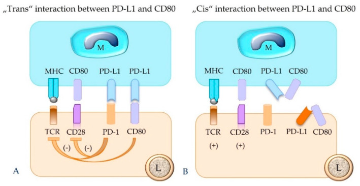
The programmed cell death protein 1/programmed death-ligand 1 (PD-1/PD-L1) pathway mainly attracted attention in immuno-oncology, leading to the development of immune checkpoint therapy. It has, however, much broader importance for tissue physiology and pathology. It mediates basic processes of immune tolerance and tissue homeostasis. In addition, it is involved in the pathogenesis of chronic infectious diseases, autoimmunity, and cancer. It is also an important paradigm for comparative pathology as well as the "one health one medicine" concept. The aim of this review is to provide an overview of novel research into the diverse facets of the PD-1/PD-L1 pathway and to give insights into its fine-tuning homeostatic role in a tissue-specific context. This review details early translational research from the discovery phase based on mice as animal models for understanding pathophysiological aspects in human tissues to more recent research extending the investigations to several animal species. The latter has the twofold goal of comparing this pathway between humans and different animal species and translating diagnostic tools and treatment options established for the use in human beings to animals and vice versa.

Keywords: PD-1; PD-L1; cancer; comparative pathology; immuno-oncology; one health one medicine concept; programmed death protein 1; programmed death protein ligand 1.


Conflict of interest statement

The authors declare no conflict of interest.

Figures

Figure 1
Figure 1
The activation state of a T cell, i.e., cytokine production, proliferation, and overall survival, is not only influenced by co-stimulation, but also by concurrent inhibitory signals. The two-signal hypothesis refers to the antigen presentation by major histocompatibility complex I or II molecules of an antigen-presenting cell that causes antigen priming of the T cell receptor (signal 1). For activation of a T cell, co-stimulation through ligation of CD80 or CD86 with CD28 is required (signal 2). The degree of the activation, however, can be further modulated by additional stimulatory or inhibitory signals. The latter are mediated by CTLA4, which competes with CD80/CD86 for CD28 binding, and by the interaction between PD-L1 and PD-1, both acting as immune checkpoint molecules. TCR = T cell receptor; MHCII = major histocompatibility complex II; CTLA4 = cytotoxic T-lymphocyte-associated protein 4; PD-1 = programmed cell death protein 1; PD-L1 = programmed death-ligand 1.
Figure 2
Figure 2
Depicted are cell populations with possible expression of the immune checkpoint molecules PD-L1 and PD-1. Notably, these molecules are upregulated by IFNγ. The binding between PD-L1 and PD-1 inhibits the functions of the PD-1-bearing target cell. DC = dendritic cell; M = macrophage; TC = tumor cell; EC = endothelial cell; Treg = regulatory T cell; Breg = regulatory B cell; B = B cell, Th1 = T helper 1 cell; Th2 = T helper 2 cell; cyTC = cytotoxic T cell; NK = natural killer cell; γδT = γδ T cell.
Figure 3
Figure 3
According to the underlying molecular mechanism, PD-L1 expression in tumor cells can be classified in four different categories, i.e., PD-L1-positive by the constitutive oncogenic pathway (A), PD-L1-positive by the adaptive induced PD-L1 expression in a “hot tumor” (B), PD-L1-negative in a “cold tumor” (C) and PD-L1-negative by the oncogenic pathway (D).
Figure 4
Figure 4
Depicted are representative areas of sections of human carcinomas that are stained with hematoxylin–eosin (AC) and immunolabeled for PD-L1 (A’C’). Immunostaining was performed with PD-L1 IHC 22C3 pharmDx for Autostainer Link 48 and DAB as chromogen. A (carcinoma of the gastro-esophageal junction): The scarce tumor stroma contains mild to moderate numbers of tumor-infiltrating lymphocytes (TILs; circle). 20×. A’: All tumor cells show linear partial to complete moderate to strong membranous staining consistent with constitutive PD-L1 expression through the oncogenic pathway. 20×. Inset: Shown in greater detail is the membrane staining of tumor cells (black arrows). In addition, a few TILs have membranous to cytoplasmic PD-L1 expression (green arrows). B (esophageal carcinoma): The tumor stroma is infiltrated with moderate TIL numbers (circles). The interface between stroma and tumor cell nests is demarcated by arrowheads. An area of necrosis is marked by asterisks. 20×. B’: PD-L1-positive immune cells accumulate at the interface between tumor cell nests and stroma (stars). The PD-L1 staining of tumor cells is restricted to those that are located at the periphery of the tumor cell nests in immediate proximity to the PD-L1-positive immune cells (hash signs). This staining pattern is consistent with adaptive induced PD-L1 expression. 20×. Inset: In higher magnification are depicted aggregates of PD-L1-positive immune cells that show a strong membranous to cytoplasmic staining (green arrows). The partial to complete membranous staining (usually of a weaker intensity compared to the adjacent immune cells) of the cancer cells is marked by black arrows. 40×. C (carcinoma of the gastro-esophageal junction): The tumor stroma is infiltrated by only a few TILs (green arrows). 20× C’: Tumor cells are PD-L1 immunonegative, whereas very few immune cells are PD-L1-positive (green arrow). 20×. Inset: The PD-L1-positive immune cell (green arrow) is depicted at higher magnification.
Figure 5
Figure 5
The “trans” interaction of PD-1 of an effector lymphocyte (L) with PD-L1 expressed by a macrophage (M), tumor cell (TC), or another lymphocyte (L) inhibits the functional activity of the PD-1-bearing target lymphocyte (L), including its survival, proliferation, and secretory activity. TCR = T cell receptor; MHC = major histocompatibility complex class I or II molecules; PD-1 = programmed cell death protein 1; PD-L1 = programmed death-ligand 1.
Figure 6
Figure 6
The binding between PD-L1 and PD-1 molecules that are expressed on the same cell (“cis” ligation) will hinder their “trans” interaction. This can result in failure of functional inhibition of the effector T cell. Depicted is “cis” interaction between PD-1 and PD-L1 in a lymphocyte (A) and a macrophage (B). M = macrophage; L = lymphocyte; TCR = T cell receptor; MHC = major histocompatibility complex class I or II molecule; PD-1 = programmed cell death protein 1; PD-L1 = programmed death-ligand 1.
Figure 7
Figure 7
In addition to the expression of PD-L1, the intrinsic expression of PD-1 has been detected on variable numbers of tumor cells from different cancer types. Therefore, PD-L1 molecules of tumor cells (TCs) can bind not only to PD-1 molecules of immune cells (ICs), but also to PD-1 molecules of tumor cells (A). Both types of cellular interactions can be blocked by anti-PD-1 antibodies (B).
Figure 8
Figure 8
Binding of the ligand PD-L1 expressed on a CD4+ Th1 cell to its receptor PD-1 present on an M1 macrophage can induce M2 trans-differentiation of the PD-1-bearing target cell (forward signaling). Subsequently, the PD-1-bearing M2 macrophage may evoke trans-differentiation of the CD4+ Th1 cell into a Th17 cell by a process named reverse signaling. Within the tumor microenvironment, such a trans-differentiation of immune cells may contribute to the shifting of an anti-cancer type I immune response to a type II immune response that is associated with an equilibrium between cancer and immune cells or cancer immune escape.
Figure 9
Figure 9
PD-L1 expressed by a macrophage (M, as depicted), tumor cell, or regulatory T cell can also bind to CD80 of a lymphocyte (L). This results in inhibition of the functional activity of the PD-1-bearing target cell (A). The “cis” interaction of PD-L1 and CD80 molecules on the same cell, e.g., a macrophage or lymphocyte, prevents their binding in “trans” and thus counteracts their inhibitory function (B). TCR = T cell receptor; MHC = major histocompatibility complex class I or II molecule; PD-1 = programmed cell death protein 1; PD-L1 = programmed death-ligand 1.

References

    1. Sundberg J.P., Schofield P.N. One medicine, one pathology, and the one health concept. J. Am. Vet. Med. Assoc. 2009;234:1530–1531. doi: 10.2460/javma.234.12.1530. - DOI - PMC - PubMed
    1. Ishida Y., Agata Y., Shibahara K., Honjo T. Induced expression of PD-1, a novel member of the immunoglobulin gene superfamily, upon programmed cell death. EMBO J. 1992;11:3887–3895. doi: 10.1002/j.1460-2075.1992.tb05481.x. - DOI - PMC - PubMed
    1. Shinohara T., Taniwaki M., Ishida Y., Kawaichi M., Honjo T. Structure, and chromosomal localization of the human PD-1 gene (PDCD1) Genomics. 1994;23:704–706. doi: 10.1006/geno.1994.1562. - DOI - PubMed
    1. Agata Y., Kawasaki A., Nishimura H., Ishida Y., Tsubata T., Yagita H., Honjo T. Expression of the PD-1 antigen on the surface of stimulated mouse T and B lymphocytes. Int. Immunol. 1996;8:765–772. doi: 10.1093/intimm/8.5.765. - DOI - PubMed
    1. Nishimura H., Nose M., Hiai H., Minato N., Honjo T. Development of lupus-like autoimmune diseases by disruption of the PD-1 gene encoding an ITIM motif-carrying immunoreceptor. Immunity. 1999;11:141–151. doi: 10.1016/S1074-7613(00)80089-8. - DOI - PubMed