. 2022 Sep 20;14(19):4553.
doi: 10.3390/cancers14194553.

Synergistic Effects of Sanglifehrin-Based Cyclophilin Inhibitor NV651 with Cisplatin in Hepatocellular Carcinoma

Affiliations

Synergistic Effects of Sanglifehrin-Based Cyclophilin Inhibitor NV651 with Cisplatin in Hepatocellular Carcinoma

Sonia Simón Serrano et al. Cancers (Basel). .

Abstract

Hepatocellular carcinoma (HCC), commonly diagnosed at an advanced stage, is the most common primary liver cancer. Owing to a lack of effective HCC treatments and the commonly acquired chemoresistance, novel therapies need to be investigated. Cyclophilins-intracellular proteins with peptidyl-prolyl isomerase activity-have been shown to play a key role in therapy resistance and cell proliferation. Here, we aimed to evaluate changes in the gene expression of HCC cells caused by cyclophilin inhibition in order to explore suitable combination treatment approaches, including the use of chemoagents, such as cisplatin. Our results show that the novel cyclophilin inhibitor NV651 decreases the expression of genes involved in several pathways related to the cancer cell cycle and DNA repair. We evaluated the potential synergistic effect of NV651 in combination with other treatments used against HCC in cisplatin-sensitive cells. NV651 showed a synergistic effect in inhibiting cell proliferation, with a significant increase in intrinsic apoptosis in combination with the DNA crosslinking agent cisplatin. This combination also affected cell cycle progression and reduced the capacity of the cell to repair DNA in comparison with a single treatment with cisplatin. Based on these results, we believe that the combination of cisplatin and NV651 may provide a novel approach to HCC treatment.

Keywords: PPIase; apoptosis; cisplatin; cyclophilin; hepatocellular carcinoma; synergy.


Conflict of interest statement

S.S.S., M.T., A.G., E.E. and M.J.H. are or have been employees of Abliva AB, which holds the commercial rights to NV651. S.S.S., A.G., E.E. and M.J.H. own shares in Abliva AB. J.M., C.K., P.G. and R.M. declare no potential conflict of interest.

Figures

Figure 3
Figure 3
Synergistic effect of NV651 and HCC treatments in HEPG2 cells. (AD) Effect on cell proliferation in HEPG2 cells. (A) HEPG2 cells were treated with NV651 in combination with sorafenib for 168 h and cell proliferation was analysed with Acumen n = 2 biological replicates. (BD) Percent inhibition of NV651 in combination with Cisplatin, Doxorubicin or Mitomycin after 72 h of treatment, analysed with resazurin. N = 1–3 biological replicates. (EH) Synergy score calculated with the highest single agent (HSA) from each data presented in (AD).
Figure 4
Figure 4
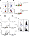
Combination effect of NV651 and Cisplatin (CDDP) on cell death. (AC) NV651’s effect on the mitochondrial membrane potential—low DiOC6(3) levels (FITC channel) correspond to low mitochondrial membrane potential (pre-apoptotic marker)—and PI+cells (upper quadrants), (late apoptotic + necrotic cells) after 72 h of exposure to the combination treatment. (A) Representative density plot of HUH7. (B,C) Quantification of PI+ cells and PIlowDiOC(6)3 cells, equivalent to the low mitochondrial membrane potential in HEPG2 (B) and HUH7 (C). (DG) SubG1 fraction from Figure 6 after combination treatment for 12, 24 and 48 h. (B,C) Total percentage of cell death statistically analysed by 2-way ANOVA followed by Dunnett’s multiple-comparison test. n = 3 biological replicates. (DF) were statistically analysed by 2-way ANOVA followed by Dunnet’s multiple comparison test, with n = 3–5 biological replicates. Data are presented as the mean ± SD * p < 0.05, ** p < 0.01 and *** p < 0.001.
Figure 5
Figure 5
Effect of NV651 and cisplatin on DNA damage for up to 24 h in drug-free media in HEPG2 and HUH7 after 4 h of pre-treatment with NV651 followed by 2 h of cisplatin exposure. Effect on DNA damage was analysed by the alkaline COMET assay. (A) Frequency distribution of the olive tail moment at 3, 6 and 24 h. (B) Representative comets at 0 and 6 h. (C,D) Percent crosslinks calculated in comparison with the control; for each sample a minimum of 50 comets were analysed. n = 3–4 biological replicates in HEPG2 (C) and n = 1 biological replicate in HUH7 (D). Data in (C) are presented as the mean ± SD.
Figure 6
Figure 6
Effects of NV651 and cisplatin on the cell cycle. DNA content was analysed with PI staining in HEPG2 and HUH7 after treatment exposure for 12, 24 or 48 h. (A) Representative histograms of HEPG2 at 48 h of exposure; (B,C) G1, S and G2/M quantification analysed by 2-way ANOVA, followed by Dunnet’s multiple comparison test, with n = 3–5 biological replicates. Data are presented as the mean ± SD * p < 0.05, ** p < 0.01 and *** p < 0.001.
Figure 1
Figure 1
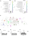
Biomarker discovery for NV651. (A) Drug efficacy prediction results using 18 signature genes in 28 grouped cell lines. The probability of being in the sensitive group is indicated as Pinsen and probability of being in the insensitive group is indicated as Pininsen. The drug response of 25 cell lines was correctly predicted. (B) Gene set enrichment analysis (GSEA) in 47 cell lines indicating the NES (normalized enrichment score), size and nominal p-value. The correlated genes were enriched in 10 pathways, with a NOM p-value of <0.01.
Figure 2
Figure 2
NV651 effect on gene expression. (AC) Transcriptome was analysed after 4 h of 500 nM NV651, CsA or control treatment in HEPG2 cells. (A,B) Top 20 genes sets for GSEA in the reactome for CsA versus NV651 (A) and control versus NV651 (B). (C) Cytoscape with the GO biological process at an intermediate level (3–8) with the significant genes indicated (kappa score ≥ 0.4). (DF) NV651’s effect on gene expression after 4 h of exposure in HEPG2 and HUH7 cells. mRNA levels of the indicated genes were analysed by qPCR. Data in (D,F) are presented as the mean ± SD of the relative expression in n = 2 biological replicates.

References

    1. World Health Organization Liver Fact Sheet. 2020. [(accessed on 1 November 2021)]. Available online: https://gco.iarc.fr/today/data/factsheets/cancers/11-Liver-fact-sheet.pdf.
    1. Janevska D., Chaloska-Ivanova V., Janevski V. Hepatocellular Carcinoma: Risk Factors, Diagnosis and Treatment. Open Access Maced. J. Med Sci. 2015;3:732–736. doi: 10.3889/oamjms.2015.111. - DOI - PMC - PubMed
    1. Raza A., Sood G.K. Hepatocellular carcinoma review: Current treatment, and evidence-based medicine. World J. Gastroenterol. 2014;20:4115–4127. doi: 10.3748/wjg.v20.i15.4115. - DOI - PMC - PubMed
    1. Llovet J.M., Di Bisceglie A.M., Bruix J., Kramer B.S., Lencioni R., Zhu A.X., Sherman M., Schwartz M., Lotze M., Talwalkar J., et al. Design and Endpoints of Clinical Trials in Hepatocellular Carcinoma. JNCI J. Natl. Cancer Inst. 2008;100:698–711. doi: 10.1093/jnci/djn134. - DOI - PubMed
    1. Tsurusaki M., Murakami T. Surgical and Locoregional Therapy of HCC: TACE. Liver Cancer. 2015;4:165–175. doi: 10.1159/000367739. - DOI - PMC - PubMed

LinkOut - more resources