CNN-Based Approaches with Different Tumor Bounding Options for Lymph Node Status Prediction in Breast DCE-MRI
- PMID: 36230497
- PMCID: PMC9558949
- DOI: 10.3390/cancers14194574
CNN-Based Approaches with Different Tumor Bounding Options for Lymph Node Status Prediction in Breast DCE-MRI
Abstract
Background: The axillary lymph node status (ALNS) is one of the most important prognostic factors in breast cancer (BC) patients, and it is currently evaluated by invasive procedures. Dynamic contrast-enhanced magnetic resonance imaging (DCE-MRI), highlights the physiological and morphological characteristics of primary tumor tissue. Deep learning approaches (DL), such as convolutional neural networks (CNNs), are able to autonomously learn the set of features directly from images for a specific task.
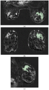
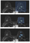
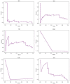
Materials and methods: A total of 155 malignant BC lesions evaluated via DCE-MRI were included in the study. For each patient's clinical data, the tumor histological and MRI characteristics and axillary lymph node status (ALNS) were assessed. LNS was considered to be the final label and dichotomized (LN+ (27 patients) vs. LN- (128 patients)). Based on the concept that peritumoral tissue contains valuable information about tumor aggressiveness, in this work, we analyze the contributions of six different tumor bounding options to predict the LNS using a CNN. These bounding boxes include a single fixed-size box (SFB), a single variable-size box (SVB), a single isotropic-size box (SIB), a single lesion variable-size box (SLVB), a single lesion isotropic-size box (SLIB), and a two-dimensional slice (2DS) option. According to the characteristics of the volumes considered as inputs, three different CNNs were investigated: the SFB-NET (for the SFB), the VB-NET (for the SVB, SIB, SLVB, and SLIB), and the 2DS-NET (for the 2DS). All the experiments were run in 10-fold cross-validation. The performance of each CNN was evaluated in terms of accuracy, sensitivity, specificity, the area under the ROC curve (AUC), and Cohen's kappa coefficient (K).
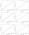
Results: The best accuracy and AUC are obtained by the 2DS-NET (78.63% and 77.86%, respectively). The 2DS-NET also showed the highest specificity, whilst the highest sensibility was attained by the VB-NET based on the SVB and SIB as bounding options.
Conclusion: We have demonstrated that a selective inclusion of the DCE-MRI's peritumoral tissue increases accuracy in the lymph node status prediction in BC patients using CNNs as a DL approach.
Keywords: axillary lymph nodes status (ALNS); bounding box; breast cancer (BC); convolutional neural network (CNN); deep learning (DL).
Conflict of interest statement
All authors submit and take responsibility for this original manuscript. All authors affirm that all contents of this manuscript have never been published or submitted for publication elsewhere. All authors approve the publication. All authors retain the copyright to the publisher.
Figures


References
-
- Rebecca L.S., Kimberly D., Miller A.J. Cancer statistics, 2018. A Cancer J. Clin. 2018;68:7–30. - PubMed
-
- Martelli G., Boracchi P., De Palo M., Pilotti S., Oriana S., Zucali R., Daidone M.G., De Palo G. A randomized trial comparing axillary dissection to no axillary dissection in older patients with t1n0 breast cancer: Results after 5 years of follow-up. Ann. Surg. 2005;242:1. doi: 10.1097/01.sla.0000167759.15670.14. - DOI - PMC - PubMed
-
- Dershaw D.D., Panicek D.M., Osborne M.P. Significance of lymph nodes visualized by the mammographic axillary view. Breast Dis. 1991;4:271–280.
Grants and funding
- no. 612462-EPP-1-2019-1-SK-EPPKA2-KA/University-Industry Educational Centre in Advanced Biomedical and Medical Informatics (CE-BMI)
- 2014-2020 CCI2014IT16M2OP005 Azione IV.4/Programma Operativo Nazionale (PON) "Ricerca e Innovazione"
- F/130096/01-05/X38/Ministero dello Sviluppo Economico (Italy) - ACCORDI PER L'INNO-VAZIONE DI CUI AL D.M. 24 MAGGIO 2017 - Fondo per la Crescita Sostenibile
LinkOut - more resources
Full Text Sources

