. 2022 Sep 24;14(19):4634.
doi: 10.3390/cancers14194634.

Treatment of Pineal Region Rosette-Forming Glioneuronal Tumors (RGNT)

Affiliations

Treatment of Pineal Region Rosette-Forming Glioneuronal Tumors (RGNT)

Anna Michel et al. Cancers (Basel). .

Abstract

Background: Rosette-forming glioneuronal tumor (RGNT) is an extremely rare entity described for the first time in the WHO classification of tumors of the central nervous system in 2007. Predominantly, single case reports of RGNT in the pineal region have been published, and specific therapy concepts are pending.

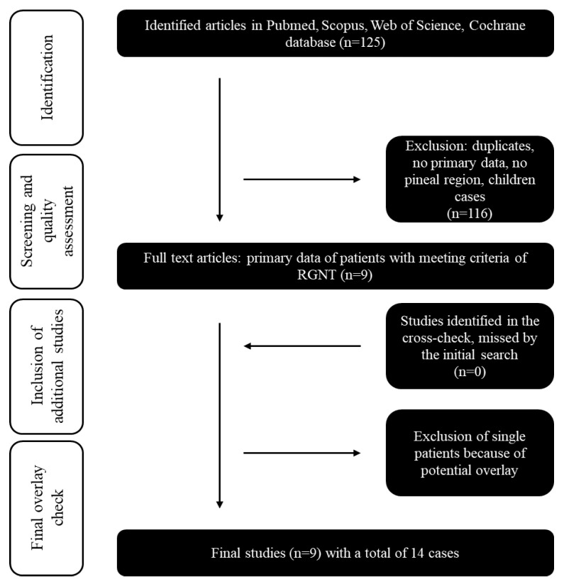
Methods: The study group comprised all patients with the RGNT (CNS WHO grade 1) in the pineal region that underwent microsurgical tumor removal in our center (August 2018-June 2021). Surgical strategy, histological findings, and clinical outcome are presented, and the results are evaluated and compared to published case reports.

Results: Four male patients aged under 50 years (range between 20 and 48 years) and one female patient, 51 years old, were included in this study. Chronic headaches and generalized epileptic seizures were the main symptoms. Supra-cerebellar infratentorial gross total tumor resection (GTR) was performed in two cases, two patients underwent subtotal tumor resection, and an endoscopic biopsy was performed in case five.

Conclusion: In cases where surgical resection seems feasible with a reasonable surgical risk, we advocate GTR. Regular and long-term MRI follow-up is essential to detect a slow tumor progression. The role of additional chemotherapy or radiotherapy remains unclear.

Keywords: RGNT; glioneuronal tumor; pineal region.


Conflict of interest statement

The authors declare no conflict of interest.

Figures

Figure 1
Figure 1
Flow chart of the systematic literature review: Inclusion and exclusion of the initial 125 studies identified by Pubmed, Scopus, Web of Science, and Cochrane databases searches. The year of publication (all cases published before 2007 were excluded) was an exclusion criterion.
Figure 2
Figure 2
The radiological findings of RGNT in the pineal region ((AD) preoperative, (A,B): T1-weighted with contrast medium, (C): T2 flair-weighted, (D): T2, (E,F): postoperative, T1-weighted with contrast medium, (G,H): 3-months follow-up, (G): T1-weighted with contrast medium, (H): T2-weighted), from the first case, a 23-year-old male patient who presented with chronic headache.
Figure 3
Figure 3
Intraoperative view before (A) and after (B) supracerebellar, infratentorial tumor resection in the semi-sitting position, exemplarily shown for case 1 (23-year-old male patient, detailed information is presented in Table 1). Abbreviations: T: tentorium; C: cerebellum; double dagger: 3rd ventricle; white arrowhead: tumor.
Figure 4
Figure 4
A benign mixed tumor with neurocytic (synaptophysin staining, (A) and astrocytic (GFAP) (F) staining, olig2 (C) staining differentiation. H&E staining (B,E) shows astrocytic differentiation (B), and typical neurocytic rosettes with perivascular pseudorosettes (E). In the Ki67 (D) staining, only very few proliferation-active cells can be recognized. (Case 1 is presented here). Abbreviations: GFAP—glial fibrillary acidic protein, H&E—haematoxylin and eosin staining.

References

    1. Konovalov A.N., Pitskhelauri D.I. Principles of treatment of the pineal region tumors. Surg. Neurol. 2003;59:250–268. doi: 10.1016/S0090-3019(03)00080-6. - DOI - PubMed
    1. Edwards M.S., Hudgins R.J., Wilson C.B., Levin V.A., Wara W.M. Pineal region tumors in children. J. Neurosurg. 1988;68:689–697. doi: 10.3171/jns.1988.68.5.0689. - DOI - PubMed
    1. Louis D.N., Perry A., Reifenberger G., von Deimling A., Figarella-Branger D., Cavenee W.K., Ohgaki H., Wiestler O.D., Kleihues P., Ellison D.W. The 2016 World Health Organization Classification of Tumors of the Central Nervous System: A summary. Acta Neuropathol. 2016;131:803–820. doi: 10.1007/s00401-016-1545-1. - DOI - PubMed
    1. Franceschi E., Frappaz D., Ruda R., Hau P., Preusser M., Houillier C., Lombardi G., Asioli S., Dehais C., Bielle F., et al. Rare Primary Central Nervous System Tumors in Adults: An Overview. Front. Oncol. 2020;10:996. doi: 10.3389/fonc.2020.00996. - DOI - PMC - PubMed
    1. Kuchelmeister K., Demirel T., Schlorer E., Bergmann M., Gullotta F. Dysembryoplastic neuroepithelial tumour of the cerebellum. Acta Neuropathol. 1995;89:385–390. doi: 10.1007/BF00309634. - DOI - PubMed

LinkOut - more resources