Review
. 2022 Sep 26;11(19):3002.
doi: 10.3390/cells11193002.

Current Understanding of the Neural Stem Cell Niches

Affiliations
Review

Current Understanding of the Neural Stem Cell Niches

Vicente Llorente et al. Cells. .

Abstract

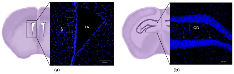
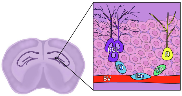
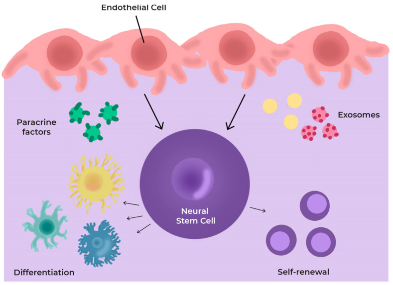
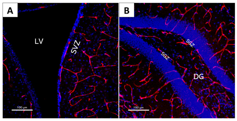
Neural stem cells (NSCs) are self-renewing, multipotent cells which give rise to all components of the central nervous system (CNS) during embryogenesis, but also activate in response to injury and disease and maintain a certain level of neurogenic activity throughout adulthood. This activity takes place in specialized regions of the brain, the neurovascular niches, whose main role is to control the behaviour of the CNS. In adult mammals, two main "canonical" niches have been described: The subventricular zone (SVZ) of the lateral ventricles and the subgranular zone (SGZ) of the dentate gyrus. This review discusses our current understanding of the neural stem cells and their canonical niches, as well as their structure, behaviours, and role in neural disease.

Keywords: brain; neurogenesis; niche; stem cell; subgranular zone; subventricular zone; vasculature.


Conflict of interest statement

The authors declare no conflict of interest. The funders had no role in the design of the study; in the collection, analyses, or interpretation of data; in the writing of the manuscript, or in the decision to publish the results.

Figures

Figure 1
Figure 1
The 20x× magnification confocal microscopy images of coronal mouse brain slices. Blue: DAPI (cell nucleus marker). (a) Detail of the subventricular zone (SVZ) within the lateral ventricle (LV), marked with arrows. (b) Detail of the subgranular zone (SGZ) within the dentate gyrus (DG), marked with arrows.
Figure 2
Figure 2
Diagram of the location and structure of the different cell types of the SVZ. LV: Lateral ventricle; E: Ependymal cells; B: Neural stem cells (NSCs), also known as radial glia-like cells (RGLs); C: Transient amplifying cells (TAPs), also known as intermediate progenitors (IPCs); A: Neuroblasts; As: Astrocytes; BV: Blood vessels.
Figure 3
Figure 3
Diagram of the location and structure of the different cell types of the SGZ. RGL: Radial glia-like cells, corresponding to NSCs and known as Type 1; IPC: Intermediate progenitor cell, also known as Type 2; NB: Neuroblast, also known as Type 3; N: Granule neuron; BV: Blood vessel, granule cell layer represented by faint pink cells in the background.
Figure 4
Figure 4
Scheme of cells and secretome in the neurovascular niche (NVN). The niche components regulate cell differentiation through the production and release of several paracrine factors and exosomes.
Figure 5
Figure 5
The 20× magnification confocal microscopy images of mouse brain slices showcasing neural stem cell niche structure and their intricate vascular network. Red: CD31 (vascular endothelial cell marker); Blue: DAPI (cell nucleus marker). (A) Lateral ventricle (LV) and subventricular zone (SVZ); (B) dentate gyrus (DG) and subgranular zone (SGZ).
Figure 6
Figure 6
Overview of potential extracellular vesicle (EV)-mediated cellular interactions in the nervous system, and the receptors or molecules involved in their integration.

References

    1. Altman J., Das G.D. Autoradiographic and histological evidence of postnatal hippocampal neurogenesis in rats. J. Comp. Neurol. 1965;124:319–335. doi: 10.1002/cne.901240303. - DOI - PubMed
    1. Reynolds B.A., Weiss S. Generation of neurons and astrocytes from isolated cells of the adult mammalian central nervous system. Science. 1992;255:1707–1710. doi: 10.1126/science.1553558. - DOI - PubMed
    1. Eriksson P.S., Perfilieva E., Björk-Eriksson T., Alborn A.M., Nordborg C., Peterson D.A., Gage F.H. Neurogenesis in the adult human hippocampus. Nat. Med. 1998;4:1313–1317. doi: 10.1038/3305. - DOI - PubMed
    1. Knoth R., Singec I., Ditter M., Pantazis G., Capetian P., Meyer R.P., Horvat V., Volk B., Kempermann G. Murine features of neurogenesis in the human hippocampus across the lifespan from 0 to 100 years. PLoS ONE. 2010;5:e8809. doi: 10.1371/journal.pone.0008809. - DOI - PMC - PubMed
    1. Boldrini M., Fulmore C.A., Tartt A.N., Simeon L.R., Pavlova I., Poposka V., Rosoklija G.B., Stankov A., Arango V., Dwork A.J., et al. Human hippocampal neurogenesis persists throughout aging. Cell Stem Cell. 2018;22:589–599. doi: 10.1016/j.stem.2018.03.015. - DOI - PMC - PubMed

Publication types

LinkOut - more resources