. 2022 Sep 29;11(19):3072.
doi: 10.3390/cells11193072.

Increased Adipose Tissue Expression of IL-23 Associates with Inflammatory Markers in People with High LDL Cholesterol

Affiliations

Increased Adipose Tissue Expression of IL-23 Associates with Inflammatory Markers in People with High LDL Cholesterol

Shihab Kochumon et al. Cells. .

Abstract

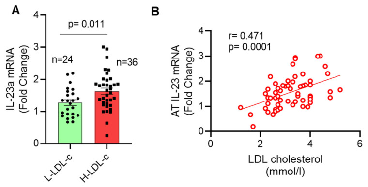
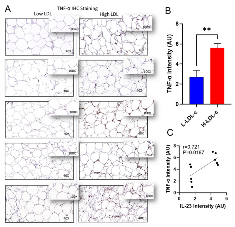
Chronic low-grade inflammation induced by obesity is a central risk factor for the development of metabolic syndrome. High low-density lipoprotein cholesterol (LDL-c) induces inflammation, which is a common denominator in metabolic syndrome. IL-23 plays a significant role in the pathogenesis of meta-inflammatory diseases; however, its relationship with LDL-c remains elusive. In this cross-sectional study, we determined whether the adipose tissue IL-23 expression was associated with other inflammatory mediators in people with increased plasma LDL-c concentrations. Subcutaneous adipose tissue biopsies were collected from 60 people, sub-divided into two groups based on their plasma LDL-c concentrations (<2.9 and ≥2.9 mmol/L). Adipose expression of IL-23 and inflammatory markers were determined using real-time qRT-PCR; plasma concentrations of total cholesterol (TC), triglyceride (TG), high-density lipoprotein cholesterol (HDL-c) and LDL-c were determined using the standard method; and adiponectin levels were measured by enzyme-linked immunosorbent assay (ELISA). Adipose IL-23 transcripts were found to be increased in people with high LDL-c, compared to low LDL-c group (H-LDL-c: 1.63 ± 0.10-Fold; L-LDL-c: 1.27 ± 0.09-Fold; p < 0.01); IL-23 correlated positively with LDL-c (r = 0.471, p < 0.0001). Immunochemistry analysis showed that AT IL-23 protein expression was also elevated in the people with H-LDL-c. IL-23 expression in the high LDL-c group was associated with multiple adipose inflammatory biomarkers (p ≤ 0.05), including macrophage markers (CD11c, CD68, CD86, CD127), TLRs (TLR8, TLR10), IRF3, pro-inflammatory cytokines (TNF-α, IL-12, IL-18), and chemokines (CXCL8, CCL3, CCL5, CCL15, CCL20). Notably, in this cohort, IL-23 expression correlated inversely with plasma adiponectin. In conclusion, adipose IL-23 may be an inflammatory biomarker for disease progression in people with high LDL-c.

Keywords: IL-23; LDL-cholesterol; adipose tissue; cytokines/chemokines; inflammation.


Conflict of interest statement

The authors declare no conflict of interest.

Figures

Figure 1
Figure 1
Increased adipose tissue IL-23 gene expression in people with high plasma LDL-c. Adipose tissue samples were obtained from 60 people and divided into two groups based on their plasma LDL-c levels as high LDL-c (<2.9 mmol/L) and low LDL-c group (≥2.9 mmol/L). Total cellular RNA was isolated from adipose tissue, and IL-23 gene expression was determined by real time qRT-PCR. Relative mRNA expression compared to GAPDH expression was presented as fold change. (A) IL-23 levels in each group are shown (bar graph). Each dot represents the individual value of IL-23, and the line represents the mean value. (B) Spearman’s correlation between IL-23 gene expression and plasma LDL-c (mmol/L) is shown. Data are represented as mean ± SEM values. Group means were compared using two-tailed unpaired Student’s t-test. A p-value of < 0.05 was considered as statistically significant.
Figure 2
Figure 2
Increased adipose tissue IL-23 protein expression in people with high LDL-c. IL-23 protein expression was assessed in people with low LDL-c (L-LDL-c) and high LDL-c (H-LDL-c) levels, 5 each, using IHC, as described in “Materials and methods”. (A) The representative IHC images obtained from three independent determinations show the elevated adipose tissue expression of IL-23 protein (40× or insert 100× magnification) in people with H-LDL-c (≥2.9 mmol/L) compared to those with L-LDL-c (˂2.9 mmol/L). (B) Increased IL-23 protein expression is shown in H-LDL-c group compared to L-LDL-c group (p < 0.0001). Staining intensity is expressed as arbitrary units (AU), determined based on Aperio-positive pixel counts (Aperio software algorithm version 9.0). IL-23 protein expression data are presented as mean ± SEM values. All p-values < 0.05 were considered statistically significant. **** Represent highly significant.
Figure 3
Figure 3
Increased adipose TNF-α protein expression in people with high LDL-c and positively correlated with IL-23 protein. TNF-α protein expression was determined in people with low LDL-c (L-LDL-c) and high LDL-c (H-LDL-c), five each, using IHC. (A) The representative IHC images obtained from three independent measurements with similar results show the elevated adipose tissue TNF-α expression (40× or insert ×100 magnification) in people with H-LDL-c (≥2.9 mmol/L) compared to those with L-LDL-c (˂2.9 mmol/L). (B) Elevated TNF-α protein expression is presented in the H-LDL-c group compared to the L-LDL-c group (p = 0.0042). Staining intensity shown as arbitrary units (AU) was determined based on Aperio-positive pixel counts (Aperio software algorithm, version 9.0). (C) IL-23 protein correlated with TNF-α protein in adipose tissue. p-values < 0.05 were considered as statistically significant. ** Highly significant.
Figure 4
Figure 4
Increased adipose CCL5 protein expression in people with high LDL-c and positively correlated with IL-23 protein. CCL5 protein expression was determined in people with low LDL-c (L-LDL-c) and high LDL-c (H-LDL-c), five each, using IHC. (A) The representative IHC images obtained from three independent determinations with similar results show the increased adipose tissue TNF-α expression (40× or inset ×100 magnification) in people with H-LDL-c (≥2.9 mmol/L) compared to those with L-LDL-c (˂2.9 mmol/L). (B) Increased CCL5 protein expression is shown in the H-LDL-c group compared to the L-LDL-c group (p = 0.0053). Staining intensity shown as arbitrary units (AU). (C) IL-23 protein correlated with CCL5 protein in adipose tissue. p-values < 0.05 were considered as statistically significant. ** Highly significant.
Figure 5
Figure 5
(A) Volcano plots represent correlation of IL-23 with differentially expressed genes in adipose tissue of people with L-LDL-c; red dots represent the significant correlation of IL-23 with other markers. (B) Volcano plots represent correlation of IL-23 with differentially expressed genes in adipose tissue of people with H-LDL-c.

References

    1. McKenzie B.S., Kastelein R.A., Cua D.J. Understanding the il-23-il-17 immune pathway. Trends Immunol. 2006;27:17–23. doi: 10.1016/j.it.2005.10.003. - DOI - PubMed
    1. Cauli A., Piga M., Floris A., Mathieu A. Current perspective on the role of the interleukin-23/interleukin-17 axis in inflammation and disease (chronic arthritis and psoriasis) Immunotargets. 2015;4:185–190. doi: 10.2147/ITT.S62870. - DOI - PMC - PubMed
    1. Gagro A., Servis D., Cepika A.M., Toellner K.M., Grafton G., Taylor D.R., Branica S., Gordon J. Type i cytokine profiles of human naive and memory b lymphocytes: A potential for memory cells to impact polarization. Immunology. 2006;118:66–77. doi: 10.1111/j.1365-2567.2006.02342.x. - DOI - PMC - PubMed
    1. Oppmann B., Lesley R., Blom B., Timans J.C., Xu Y., Hunte B., Vega F., Yu N., Wang J., Singh K., et al. Novel p19 protein engages il-12p40 to form a cytokine, il-23, with biological activities similar as well as distinct from il-12. Immunity. 2000;13:715–725. doi: 10.1016/S1074-7613(00)00070-4. - DOI - PubMed
    1. Teng M.W., Bowman E.P., McElwee J.J., Smyth M.J., Casanova J.L., Cooper A.M., Cua D.J. Il-12 and il-23 cytokines: From discovery to targeted therapies for immune-mediated inflammatory diseases. Nat. Med. 2015;21:719–729. doi: 10.1038/nm.3895. - DOI - PubMed

Publication types

MeSH terms