Review
. 2022 Sep 30;11(19):3079.
doi: 10.3390/cells11193079.

Technologies Enabling Single-Molecule Super-Resolution Imaging of mRNA

Affiliations
Review

Technologies Enabling Single-Molecule Super-Resolution Imaging of mRNA

Mark Tingey et al. Cells. .

Abstract

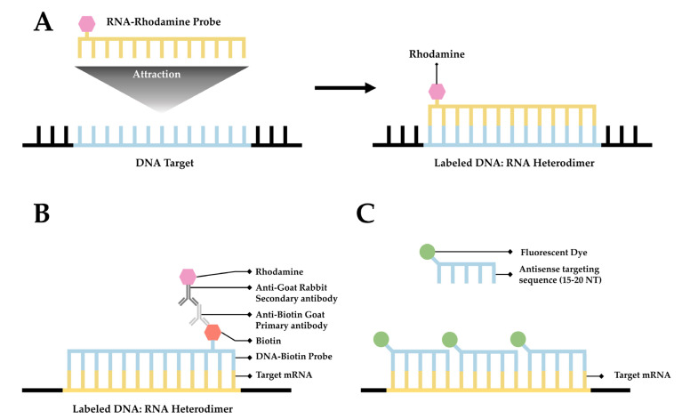
The transient nature of RNA has rendered it one of the more difficult biological targets for imaging. This difficulty stems both from the physical properties of RNA as well as the temporal constraints associated therewith. These concerns are further complicated by the difficulty in imaging endogenous RNA within a cell that has been transfected with a target sequence. These concerns, combined with traditional concerns associated with super-resolution light microscopy has made the imaging of this critical target difficult. Recent advances have provided researchers the tools to image endogenous RNA in live cells at both the cellular and single-molecule level. Here, we review techniques used for labeling and imaging RNA with special emphases on various labeling methods and a virtual 3D super-resolution imaging technique.

Keywords: CRISPR-Cas13; CRISPR-Cas9; FISH; MS2-MCP; MTRIPs; SMLM; mRNA; molecular beacons; seqFISH; single-molecule super-resolution microscopy; smFISH.


Conflict of interest statement

The authors declare no conflict of interest.

Figures

Figure 1
Figure 1
A simplified diagram depicting fluorescent in situ hybridization (FISH). (A) The first FISH experiment in DNA performed by Bauman and colleagues [52], in which a target DNA sequence (Blue) forms and RNA:DNA heterodimer with an RNA (Green) probe conjugated with Rhodamine (Red). (B) The first mRNA FISH experiment performed by Singer & Ward [54], in which a target mRNA sequence (Green) formed a DNA:RNA heterodimer with a complimentary DNA sequence (Blue) conjugated to biotin (purple). A primary anti-Biotin Goat derived primary antibody (Dark Blue) associates with the biotin tag. A secondary anti-Goat rabbit derived antibody (Light Red) conjugated to a Rhodamine (Dark Red) then associates with the primary antibody forming a complete fluorescent label. (C) The core principle of single-molecule FISH (smFISH), in which a target mRNA (Green) is targeted with short sequential antisense oligonucleotides (Blue), each 15–20 nucleotides long, that are each conjugated to a fluorescent dye (Dark Green).
Figure 2
Figure 2
smFISH and seqFISH. (A) smFISH: Top, multiple probes of the same color designed to hybridize along the length of both the CDS and 3′UTR of the intended transcript; bottom, mRNAs imaged after hybridization, with yellow spots showing transcripts containing both the CDS and 3′UTR [after (Raj, 2008)]. (B) seqFISH Spatial barcoding, in which probes are designed to hybridize ~100 nt apart to facilitate resolution of unique combinations (after [71]). (C) Spectral barcoding, in which a color code of probes hybridizes repeatedly along the length of a transcript in order to increase its detectability and identifiability. (D) Repeated cycles of hybridization, imaging, and removal of probes results in a temporal barcode that increases the number of unique barcodes possible and aids resolution via the temporal dimension (Panels C,D after [73]). © seqFISH in vitro: An oligo(dT) surface is created and mRNAs hybridize to it via their poly-A tail, spreading out to a resolvable distance. (F) Probes are hybridized to the adhered transcripts.
Figure 3
Figure 3
The MCP-MS2 loop system. (A) A depiction of the gene cassettes present in the two plasmids, the chimeric target sequence and the MCP-fluorophore, utilized in this system. (B) A simplified diagram of the association between the chimeric target sequence and the MCP-Fluorophore post transcription/translation. (C) A depiction of the fluorescent pattern observed in cells that have only the MCP-Fluorophore plasmid. (D) A depiction of the fluorescent pattern observed in cells that contain both the MCP-Fluorophore and Chimeric Target Sequence plasmids.
Figure 4
Figure 4
Simplified schematic of MTRIPs. (A) Fluorophores are bound to synthetic oligomers (ligands). (B) Tagged ligands are introduced into the live cell via temporary permeabilization. (C) Ligands bind to target mRNA (not o scale); multiply bound mRNAs are distinguished from unbound probes by intensity. After [123].
Figure 5
Figure 5
Nucleocytoplasmic transportation and mRNA imaging utilizing CRISPR. CRISPR/Cas9 is guided to DNA via sgRNA and neighboring PAM. Nuclease activity of Cas9 makes a double-stranded break, allowing for insertion of targeting aptamers such as MS2 hairpin loops, and for a fluorescent protein fused to MCP to label the modified mRNA. A nuclease deficient Cas (dCas) protein fused with a fluorescent protein can be used to bind to mRNA of interest directly instead, without genetic modification. By providing a double-stranded PAMmer with dCas9, dCas9 can be made to bind with mRNA instead of DNA. Alternatively, dCas13 natively binds to mRNA without the need for PAMmer sequence.
Figure 6
Figure 6
Simplified diagram of RNA molecular beacons. (A) A 15- to 20-nucleotide target sequence flanked by palindromic repeats causes the probe to form a stem-loop, bringing the quencher (Grey) and a fluorophore into close proximity causing the fluorophore to quench (Dark Grey). (B) When in close proximity to the target transcript, the target sequence will hybridize with the target mRNA, causing the stem-loop to open, moving the quencher and fluorophore away from one another, thereby facilitating fluorescence.

References

    1. Crick F. Central dogma of molecular biology. Nature. 1970;227:561–563. doi: 10.1038/227561a0. - DOI - PubMed
    1. Noller H.F. Ribosomal RNA and translation. Annu. Rev. Biochem. 1991;60:191–227. doi: 10.1146/annurev.bi.60.070191.001203. - DOI - PubMed
    1. Elela S.A., Nazar R.N. Role of the 5.8 S rRNA in ribosome translocation. Nucleic Acids Res. 1997;25:1788–1794. doi: 10.1093/nar/25.9.1788. - DOI - PMC - PubMed
    1. Dana H., Chalbatani G.M., Mahmoodzadeh H., Karimloo R., Rezaiean O., Moradzadeh A., Mehmandoost N., Moazzen F., Mazraeh A., Marmari V. Molecular mechanisms and biological functions of siRNA. Int. J. Biomed. Sci. IJBS. 2017;13:48. - PMC - PubMed
    1. Moore C.B., Guthrie E.H., Huang M.T., Taxman D.J. Short hairpin RNA (shRNA): Design, delivery, and assessment of gene knockdown. Methods Mol. Biol. 2010;629:141–158. doi: 10.1007/978-1-60761-657-3_10. - DOI - PMC - PubMed

Publication types

MeSH terms

LinkOut - more resources