. 2022 Oct 5;11(19):3129.
doi: 10.3390/cells11193129.

FGFR2 Mutation p.Cys342Arg Enhances Mitochondrial Metabolism-Mediated Osteogenesis via FGF/FGFR-AMPK-Erk1/2 Axis in Crouzon Syndrome

Affiliations

FGFR2 Mutation p.Cys342Arg Enhances Mitochondrial Metabolism-Mediated Osteogenesis via FGF/FGFR-AMPK-Erk1/2 Axis in Crouzon Syndrome

Yidi Wang et al. Cells. .

Abstract

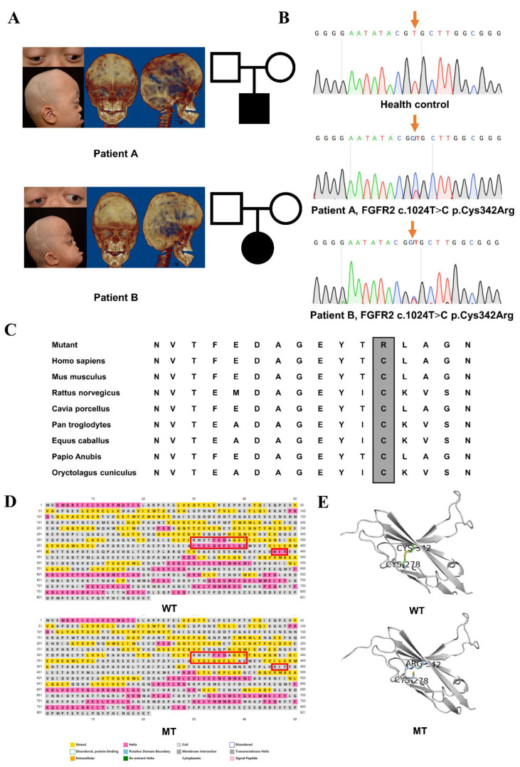
Background: Crouzon syndrome ([OMIM] #123500) caused by FGFR2 mutation is an autosomal dominant syndrome with craniosynostosis, the underlying mechanism of which remains obscure.

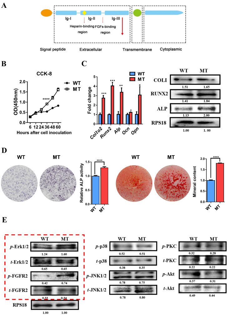
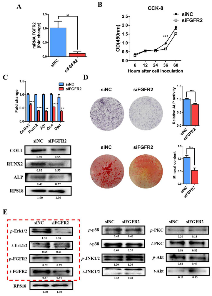
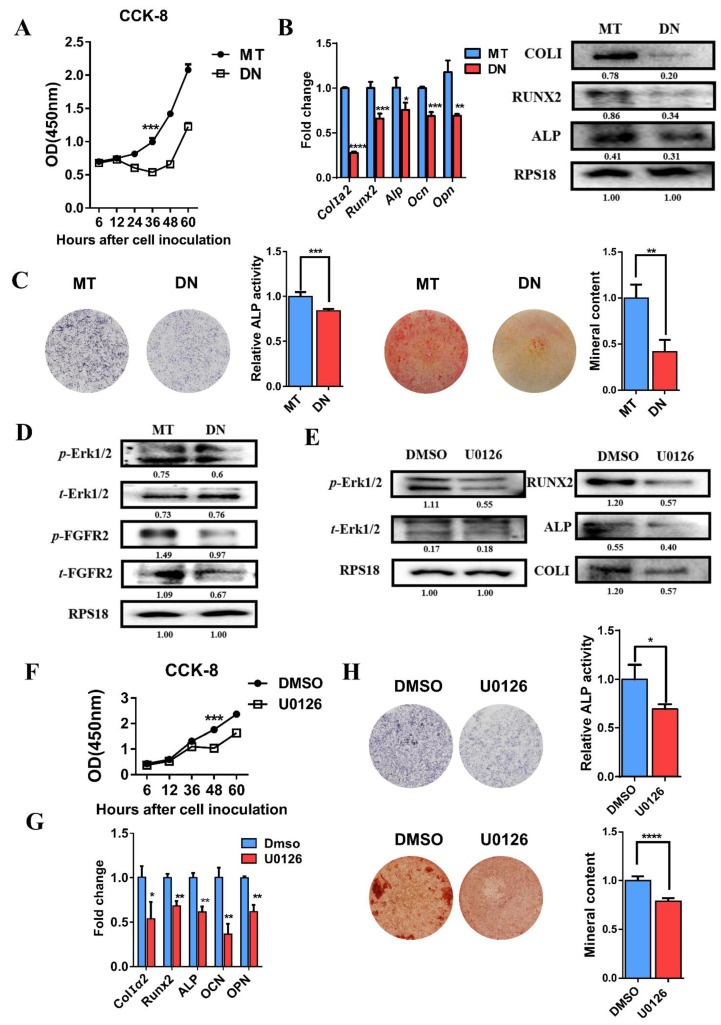
Methods: First, whole exome sequencing was used to screen the possible pathogenic variant in two sporadic patients with Crouzon syndrome. The investigation of primary and secondary structures as well as the conservation analysis of FGFR2 mutation (p.Cys342Arg) was performed. Then, wild-type and mutant overexpression plasmids were constructed and transfected into pre-osteoblastic murine cell line MC3T3-E1 cells. Osteogenesis and mitochondrial metabolism were analyzed by CCK8, ALP staining and ALP activity, alizarin red staining, qRT-PCR, Western blot, seahorse assays and mitochondrial staining. The siRNA targeting FGFR2 and domain negative FGFR2 were designed for verification.

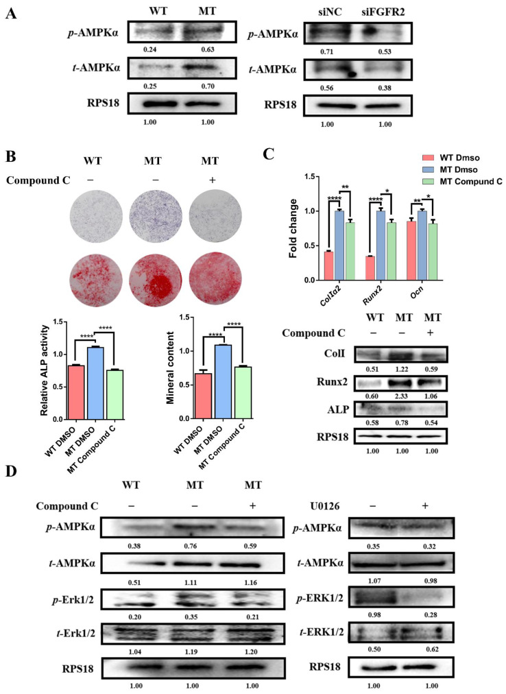
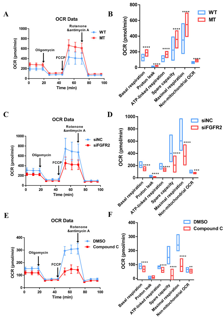
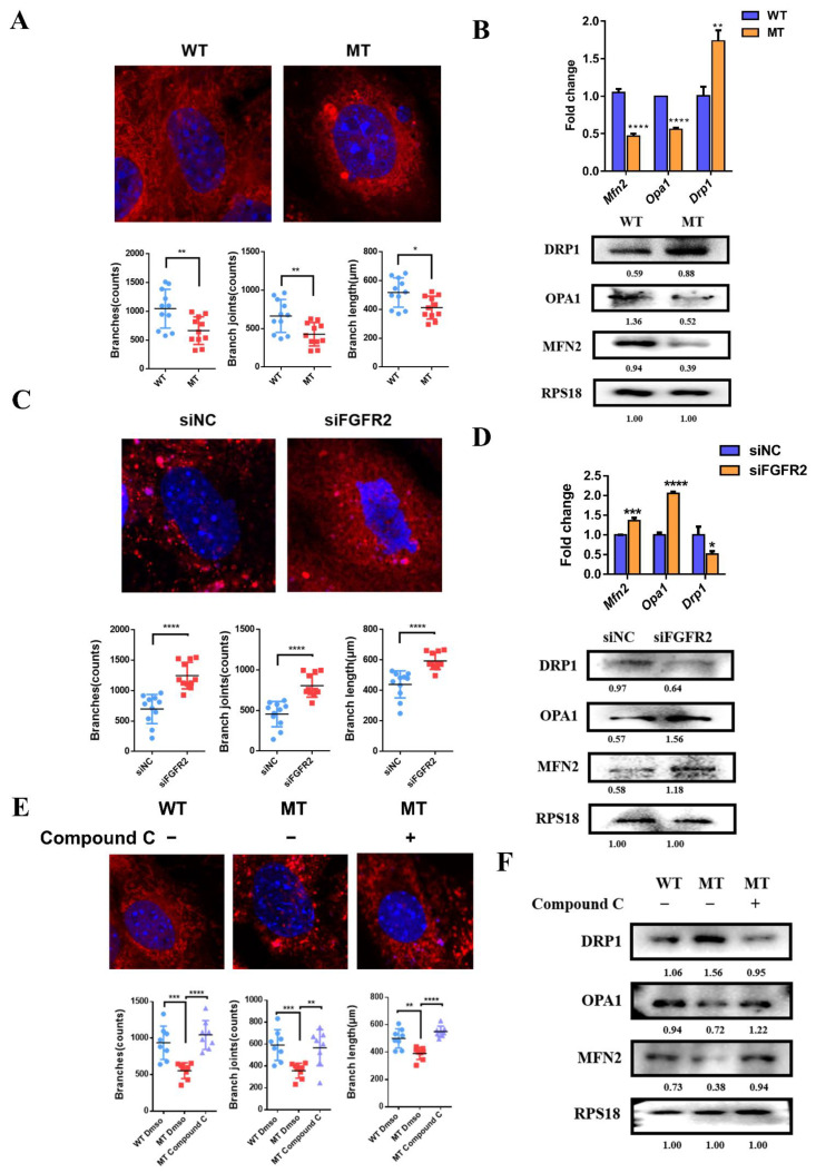
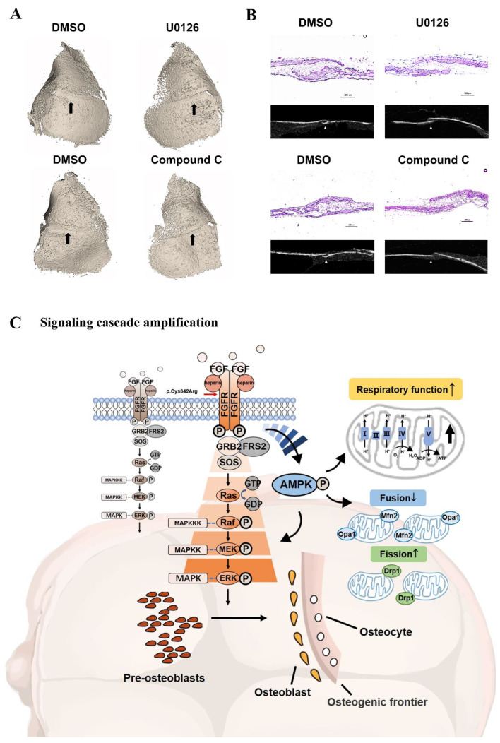
Results: First, FGFR2 mutation (p.Cys342Arg) was detected in two sporadic Chinese Crouzon syndrome patients. FGFR2 p.Cys342Arg promoted the osteogenic differentiation of MC3T3-E1 cells through the upregulation of AMP-activated protein kinase (AMPK)-Erk1/2 signal pathway. Furthermore, FGFR2 p.Cys342Arg enhanced oxidative phosphorylation and converted mitochondrial fusion to the fission of MC3T3-E1, promoting osteogenic differentiation and craniosynostosis in Crouzon syndrome. Additionally, AMPK or Erk1/2 inhibitors delayed the cranial suture closure.

Conclusion: FGFR2 mutation p.Cys342Arg promotes osteogenesis by enhancing mitochondrial metabolism-mediated via FGF/FGFR-AMPK-Erk1/2 axis, which indicates the potential of therapy targeting AMPK or Erk1/2 for syndromic craniosynostosis treatment.

Keywords: AMPK; constitutive activation mutation; craniosynostosis; mitochondrial metabolism; osteogenic differentiation.


Conflict of interest statement

The authors declare no potential conflicts of interest with respect to the authorship and/or publication of this article.

Figures

Figure 1
Figure 1
Clinical findings, identified mutation and pathogenic assessments: (A) Digital photos and CT of two sporadic Chinese children with Crouzon syndrome after cranial vault reconstruction. Both of them suffered from the premature closure of the bilateral coronal sutures, acrobrachycephaly, exophthalmos, midfacial hypoplasia and crossbite. (B) Sequencing chromatograms of the identified FGFR2 c.1024T>C mutation which was marked by an orange arrow and wild-type sequence. (C) Residue at codon 342, indicated by the frame, was evolutionarily conserved among all tested species. (D) Helices were reduced in p.Cys342Arg mutant FGFR2 circled by red boxes. (E) A disulfide bond which was originally formed by cysteine at codon 342 and cysteine at codon 278 was disconnected because of p.Cys342Arg.
Figure 2
Figure 2
Constitutive activation of FGFR2 (p.Cys342Arg) enhanced osteogenesis via Erk1/2 MAPK signaling pathway: (A) Schematic representation of the relative linear location in which the FGFR2 mutation is identified as illustrated by the large red arrow shown in the context of the protein structure. (B) CCK-8 assay was carried out to assess cell proliferation. Proliferation of MC3T3-E1 cells was more active in the MT group. (C) Relative expressions of osteogenic marker measured by qPCR. The expressions of Alp, ColIα2, Runx2, Opn and Ocn mRNA in differentiated MC3T3-e1 were remarkably increased in the MT group. Western blot analysis showed that the level of ALP, COLI and RUNX2 were also increased in the MT group. (D) ALP staining, alizarin red staining and quantitative tests. ALP staining showed increased crystal violet-staining cells in the MT group compared with the WT group. Quantitative experiment demonstrated that ALP activity is more active in the MT group. Alizarin red staining and quantitative test showed there were more mineralized nodules and mineral content in the MT group than in the WT group. (E) Western blot analysis demonstrated that the levels of p-FGFR2 and p-Erk1/2 were increased in the MT group. There was no significant change in the expression of key proteins in other downstream pathways. The western blot results of FGF/FGFR2-Erk1/2 was circled by the red frame. p values were significant at * p < 0.05, ** p < 0.01, *** p < 0.001 and **** p < 0.0001.
Figure 3
Figure 3
Knockdown of FGFR2 induced by siRNA-inhibited osteogenic differentiation by decreasing Erk1/2 MAPK activation: (A) qRT-PCR assay showed mRNA FGFR2 expression after transfection with siRNA against FGFR2. (B) CCK-8 assays were carried out to assess cell proliferation. Knockdown of FGFR2 inhibited the proliferation of MC3T3-E1 cells. (C) Relative expressions of osteogenic marker measured by qRT-PCR and Western blot. The expression of Alp, ColIα2, Runx2, Opn and Ocn mRNA in differentiated MC3T3-E1 were remarkably decreased in siFGFR2 group. The level of ALP, COLI, RUNX2 protein were also decreased in siFGFR2 group. (D) ALP staining and quantitation of ALP activity showed that the ALP activity was decreased in the siFGFR2 group compared with the control. Alizarin red staining and quantitative test demonstrated there was less mineral content in the siFGFR2 group. (E) Western blot analysis demonstrated that the level of t-FGFR2, p-FGFR2 and p-Erk1/2 was decreased in the siFGFR2 group. There was no significant change in the expression of key proteins in other downstream pathways. The western blot results of FGF/FGFR2-Erk1/2 was circled by the red frame. p values were significant at ** p < 0.01, *** p < 0.001 and **** p < 0.0001.
Figure 4
Figure 4
Domain negative FGFR2 weakened osteogenesis through the Erk1/2 signaling pathway: (A) CCK-8 assays showed the proliferation of MC3T3-E1 cells was inhibited in the DN FGFR2 group compared with the MT group. (B) Relative expressions of osteogenic genes such as ColIα2, Runx2, Alp, Ocn and Opn were decreased in the DN group compared with the MT group measured by qRT-PCR. The level of ALP, COLI and RUNX2 was inhibited in the DN group, as detected by Western blot. (C) ALP staining and quantification of the ALP activity of MC3T3-E1 was decreased in the DN group. Alizarin red staining and the quantification of mineral content demonstrated that the mineral content of MC3T3-E1 was significantly reduced in the DN group. (D) Western blot showed that the expressions of t-FGFR2, p-FGFER2 and p-Erk1/2 were remarkably decreased in the DN group. (E) Alterations after using U0126 proved the positive role of Erk1/2 MAPK on osteogenesis. Western blot demonstrated that the Erk1/2 MAPK pathway was inhibited by U0126 and the levels of osteogenic marker, COLI, RUNX2 and ALP were decreased. (F) MEK inhibitor, U0126, suppressed the proliferation of MC3T3-E1 cells. (G) The expressions of Alp, ColIα2, Runx2, Opn and Ocn mRNA detected by qRT-PCR were reduced by using U0126. (H) ALP staining, alizarin red staining and quantification test demonstrated that both ALP activity and mineralization were decreased after the Erk1/2 MAPK pathway was inhibited. p values were significant at * p < 0.05, ** p < 0.01, *** p < 0.001 and **** p < 0.0001.
Figure 5
Figure 5
The activation of the AMPK-Erk pathway contributed to dysregulating osteogenesis by FGFR2 p.Cys342Arg: (A) Western blot analysis showed that the expression of t-AMPK and p-AMPK were upregulated in the MT group compared with the WT group and the expression of t-AMPK and p-AMPK were downregulated in the siFGFR2 group compared with the control. (B) ALP staining, alizarin red staining and quantification test demonstrated that both ALP activity and mineralization were decreased after the AMPK pathway was inhibited. (C) The expressions of osteogenic markers Alp, ColI, Runx2 and Ocn were reduced after being treated with Compound C, as determined by qRT-PCR and Western blot. (D) Western blot analysis showed that the level of p-Erk was effectively inhibited after being treated with Compound C. However, the level of p-AMPK had no significant change after the U0126 treatment. p values were significant at * p < 0.05, ** p < 0.01 and **** p < 0.0001.
Figure 6
Figure 6
Mitochondria respiratory dysfunction influenced by the FGFR/FGFR2-AMPK pathway: (A) Traces of OCR in the WT group and MT group, respectively, in response to oligomycin, FCCP, and rotenone and antimycin A. (B) All parameters of cell respiratory function in the MT group were enhanced to varying degrees. The maximal respiratory capacity was significantly higher in the MT group than that in the WT group—547.61 ± 87.60 pmol/min vs. 350.64 ± 109.08 pmol/min, respectively. The same was true for spare capacity, with 358.39 ± 64.05 pmol/min in the MT group and 222.63 ± 84.41 pmol/min in the WT group. (C) Traces of OCR in the siNC group and siFGFR2 group. (D) After the knockdown of FGFR2, a completely opposite trend revealed itself. The maximal respiration (354.50 ± 95.67 pmol/min) and spare capacity (209.24 ± 78.07 pmol/min) were about the half of maximal respiration (605.74 ± 161.03 pmol/min) and spare capacity (398.69 ± 135.26 pmol/min) in the siNC group. (E) OCR traces were then treated with Compound C, an AMPK inhibitor. DMSO was added as the control. (F) Indicators of cell respiratory function were decayed, especially maximal OCR (20.70 ± 20.95 pmol/min) and spare respiratory capacity (86.59 ± 21.92 pmol/min), compared to 150.93 ± 32.84 pmol/min of maximal OCR and 239.42 ± 35.41 pmol/min of spare respiratory capacity in the control. p values were significant at ** p < 0.01, *** p < 0.001 and **** p < 0.0001.
Figure 7
Figure 7
Mitochondrial dynamics participate in osteogenesis mediated by FGFR/FGFR2-AMPK pathway: (A) Mito-tracker red and mitochondria 2D analysis showed more fragmented or punctate mitochondria with fewer and shorter branches in the MT group, while there were widely reticular mitochondria with longer branches in the WT group. (B) qRT-PCR and Western blot analysis demonstrated that the expressions of mitochondrial-fusion-related factors Mfn2 and Opa1 were downregulated, however, the expression of mitochondrial-fission-related factor Drp1 was upregulated in the MT group compared to the WT group. (C) Mito-tracker red and mitochondria 2D analysis showed a filamentous network of mitochondria with more and longer branches widely spread in the cytoplasm in the siFGFR2 group. (D) qPCR and Western blot analysis showed that the expression of fusion-related genes Mfn2 and Opa1 was increased and the expression of fission-related gene Drp1 was decreased in the siFGFR2 group. (E) After treatment with Compound C, the morphology of the mitochondria which should have tended to split adopted a fused reticular structure with more and longer branches, as shown by Mito-tracker red and mitochondria 2D analysis. (F) Western blot analysis showed the level of MFN2 and OPA1 was increased and then level of DRP1 was decreased, as induced by Compound C. p values were significant at * p < 0.05, ** p < 0.01, *** p < 0.001 and **** p < 0.0001.
Figure 8
Figure 8
(A) U0126 and Compound C attenuated the closure of coronal sutures of cultured calvarias. It was shown by 3D reconstruction images that there was an obvious linear gap between the frontal and parietal bone after the treatment with U0126 and Compound C. In the control groups, dense bony unions were formed in coronal suture regions. The coronal suture region was marked by a black arrow. (B) As shown by the section selected at the corresponding position of micro-CT, there were gaps between the osteogenic fronts of frontal and parietal bone in the inhibitor groups. The osseous cross-linking in the coronal suture was closer in control groups. As revealed by H&E staining, the synostosis between frontal and parietal bone presented as serrated in the control. However, there was a decreased overlapping region after treatment with U0126 and Compound C. (C) Schematic diagram of osteogenesis and internal mechanism. In the process of pre-osteoblast cells further differentiating, the relatively complete FGF/FGFR2-AMPK-Erk1/2 MAPK pathway plays a pivotal role. The constitutive activation of the AMPK-Erk1/2 signaling path network leads to enhanced osteogenesis because of the FGFR2 mutation (p.Cys342Arg) differing from wild-type FGFR2. In addition to being upstream of the Erk1/2 signal, the expression and activation of AMPK amplifies the mitochondria respiratory function and changes the ratio of mitochondrial division and fusion to respond to osteogenic activity. The differentiated osteoblasts then secrete the extracellular matrix and subsequently promote mineral deposition at the front edge of osteogenesis, which may lead to the malformation of cranial vault bones in Crouzon syndrome.

References

    1. Mitulla B., Hinkel G.K., Lorenz P. [Crouzon syndrome (Mc K 12350)] Kinderarztl. Prax. 1991;59:278–280. - PubMed
    1. Wang J.C., Nagy L., Demke J.C. Syndromic Craniosynostosis. Facial Plast. Surg. Clin. N. Am. 2016;24:531–543. doi: 10.1016/j.fsc.2016.06.008. - DOI - PubMed
    1. Lin Y., Gao H., Ai S., Eswarakumar J.V.P., Zhu Y., Chen C., Li T., Liu B., Jiang H., Liu Y., et al. FGFR2 mutations and associated clinical observations in two Chinese patients with Crouzon syndrome. Mol. Med. Rep. 2017;16:5841–5846. doi: 10.3892/mmr.2017.7397. - DOI - PMC - PubMed
    1. Wilkie A.O., Bochukova E.G., Hansen R.M., Taylor I.B., Rannan-Eliya S.V., Byren J.C., Wall S.A., Ramos L., Venancio M., Hurst J.A., et al. Clinical dividends from the molecular genetic diagnosis of craniosynostosis. Am. J. Med. Genet. A. 2007;143A:1941–1949. doi: 10.1002/ajmg.a.31905. - DOI - PubMed
    1. Bonaventure J., El Ghouzzi V. Molecular and cellular bases of syndromic craniosynostoses. Expert. Rev. Mol. Med. 2003;5:1–17. doi: 10.1017/S1462399403005751. - DOI - PubMed

Publication types

MeSH terms

Substances

LinkOut - more resources