. 2022 Sep 22;23(19):11170.
doi: 10.3390/ijms231911170.

Anti-TNF Therapies Suppress Adipose Tissue Inflammation in Crohn's Disease

Affiliations

Anti-TNF Therapies Suppress Adipose Tissue Inflammation in Crohn's Disease

Albert Boronat-Toscano et al. Int J Mol Sci. .

Abstract

Anti-TNF biologics have been shown to markedly improve the quality of life for patients with Crohn's disease (CD), yet one-third of patients fail to benefit from this treatment. Patients with CD develop a characteristic wrapping of visceral adipose tissue (VAT) in the inflamed intestinal area, termed creeping fat, and it is known that adipose tissue expansion influences the efficacy of anti-TNF drugs. We questioned whether anti-TNF therapies impact the creeping fat in CD, which might affect the outcome of the disease. Adipose tissue biopsies were obtained from a cohort of 14 patients with CD that received anti-TNF drugs and from 29 non-anti-TNF-treated patients (control group) matched by sex, age, and body mass index undergoing surgical interventions for symptomatic complications. We found that anti-TNF therapies restored adipose tissue morphology and suppressed immune cell infiltration in the creeping fat. Additionally, anti-TNF treatments appeared to markedly improve the pro-inflammatory phenotype of adipose-tissue macrophages and adipose-tissue-derived stem cells. Our study provides evidence that anti-TNF medications influence immune cells and progenitor cells in the creeping of patients with CD, suppressing inflammation. We propose that perilesional VAT should be considered when administering anti-TNF therapy in patients with CD.

Keywords: TNF; adalimumab; adipose tissue; creeping fat; infliximab.


Conflict of interest statement

The authors declare no conflict of interest.

Figures
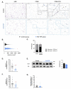
Figure 1
Figure 1
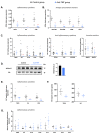
Anti-TNF therapy remodels adipose tissue, restoring normal adipocyte size and suppressing immune cell infiltration. (A) Representative histological images obtained from randomly selected 304,558-µm2 circular sections. Left panel: hematoxylin & eosin (H&E) staining. Central panel: CD68 immunohistochemistry (brown staining). Right panel: double CD68 (magenta stain) and CD3 immunohistochemistry (brown stain). (B) Adipocyte area in the creeping fat of control group and anti-TNF-treated group. (C) Percentage of large-size (>3000 µm2) and small-size (<3000 µm2) adipocytes detected in creeping fat. (D) Number of CD68-positive macrophages per section detected in creeping fat. (E) CD14 protein expression analyzed by Western blotting in creeping fat of control and anti-TNF-treated group. (F) Number of CD3-positive T lymphocytes in the creeping fat of control and anti-TNF-treated group. (G) Ratio of CD68- to CD3-positive cells in creeping fat. Results are shown as mean ± SEM from independent donors’ experiments. * p < 0.05, *** p < 0.001, and **** p < 0.0001 vs. control group.
Figure 2
Figure 2
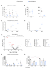
Anti-TNF treatment suppresses inflammation in different cell subtypes of creeping fat. (A) Gene expression of pro-inflammatory genes (TNFA, IL6, and IL1B) in VAT-derived adipose tissue macrophages (ATMs). (B) Gene expression of antigen-presentation markers in VAT-derived ATMs. (C) Gene expression of the inflammatory cytokines TNFA, IL6, IL1B, CCL2, and IL12B; anti-inflammatory cytokines TGFB1, IL10, and ADIPOQ; the invasion markers MMP2 and MMP9 in the creeping fat of patients treated with anti-TNF or control group. (D) NFKB-p52 protein expression analyzed by Western blotting in the creeping fat of patients treated with anti-TNF or control group (E) Gene expression of the pro-inflammatory cytokines TNFA, IL6, and IL1B in VAT explants isolated from patients treated with anti-TNF or control group. (F) TNFA and IL6 abundance in 24 h conditioned medium of VAT explants from patients treated with anti-TNF or control group. (G) Inflammatory cytokine gene expression in mature adipocytes isolated from patients treated with anti-TNF or control group. Results are shown as mean ± SEM from independent donors’ experiments. * p < 0.05 and ** p < 0.01 vs. control group.
Figure 3
Figure 3
Anti-TNF therapies modify the inflammatory status of creeping fat-derived ASCs. (A) Gene expression of pro-inflammatory genes (TNFA, IL6, IL1B, and CCL2) in adipose-stem cells (ASCs) and (B) TNFA and IL6 abundance in 24 h conditioned medium of ASCs from patients treated with anti-TNF or control group. (C) Gene expression of anti-inflammatory genes (IL10, CGSF, and TGFB1) in ASCs from patients treated with anti-TNF or control group. (D) TGFB1 protein abundance in 24 h conditioned medium of ASCs from patients treated with anti-TNF or control group. (E) Analysis of the ASC protein secretome and protein abundance differences between ASCs isolated from control group (n = 6) and anti-TNF-treated group (n = 3). (F) Gene expression of antigen-presentation markers (HLA-DM and HLA-DPB) and co-stimulatory molecules (CD40, CD74, and CD80) in ASCs from patients treated with anti-TNF or control group. (G) ASCs isolated from patients treated with anti-TNF or control group co-cultured or not with ovalbumin (OVA) were stained with a panel of antibodies and analyzed by flow cytometry to compare the percentage of surface expression of antigen-presenting markers. Results are shown as mean ± SEM from independent donors’ experiments. * p < 0.05 vs. control group. $ p < 0.05 control group without OVA vs. control group with OVA.

References

    1. Sarmento A. Editorial: Understanding Crohn’s Disease: Immunity, Genes and Microbes. Front. Immunol. 2017;8:357. doi: 10.3389/fimmu.2017.00357. - DOI - PMC - PubMed
    1. Büning C., von Kraft C., Hermsdorf M., Gentz E., Wirth E.K., Valentini L., Haas V. Visceral Adipose Tissue in Patients with Crohn’s Disease Correlates with Disease Activity, Inflammatory Markers, and Outcome. Inflamm. Bowel. Dis. 2015;21:2590–2597. doi: 10.1097/MIB.0000000000000527. - DOI - PubMed
    1. Connelly T.M., Juza R.M., Sangster W., Sehgal R., Tappouni R.F., Messaris E. Volumetric Fat Ratio and Not Body Mass Index Is Predictive of Ileocolectomy Outcomes in Crohn’s Disease Patients. Dig. Surg. 2014;31:219–224. doi: 10.1159/000365359. - DOI - PubMed
    1. Li X.-H., Feng S.-T., Cao Q.-H., Coffey J.C., Baker M.E., Huang L., Fang Z.-N., Qiu Y., Lu B.-L., Chen Z.-H., et al. Degree of Creeping Fat Assessed by Computed Tomography Enterography Is Associated with Intestinal Fibrotic Stricture in Patients with Crohn’s Disease: A Potentially Novel Mesenteric Creeping Fat Index. J. Crohns. Colitis. 2021;15:1161–1173. doi: 10.1093/ecco-jcc/jjab005. - DOI - PMC - PubMed
    1. Kredel L.I., Siegmund B. Adipose-Tissue and Intestinal Inflammation—Visceral Obesity and Creeping Fat. Front. Immunol. 2014;5:462. doi: 10.3389/fimmu.2014.00462. - DOI - PMC - PubMed