. 2022 Sep 26;23(19):11347.
doi: 10.3390/ijms231911347.

Interspecies Comparison of Interaction Energies between Photosynthetic Protein RuBisCO and 2CABP Ligand

Affiliations

Interspecies Comparison of Interaction Energies between Photosynthetic Protein RuBisCO and 2CABP Ligand

Masayasu Fujii et al. Int J Mol Sci. .

Abstract

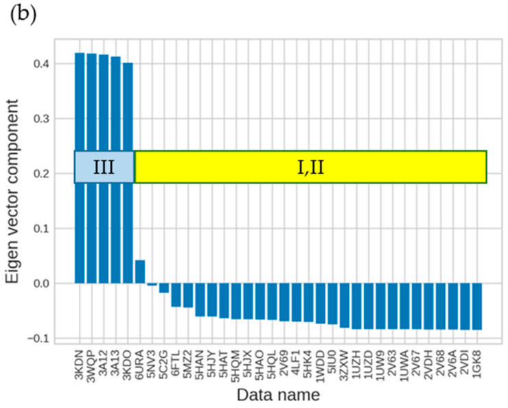
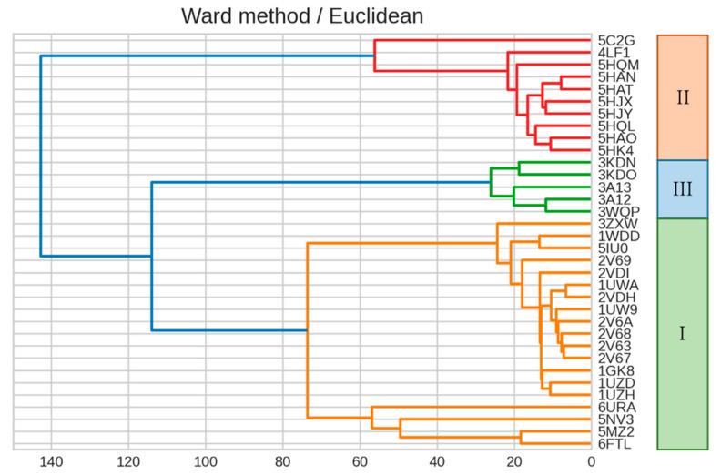
Ribulose 1,5-bisphosphate carboxylase/oxygenase (RuBisCO) functions as the initial enzyme in the dark reactions of photosynthesis, catalyzing reactions that extract CO2 from the atmosphere and fix CO2 into organic compounds. RuBisCO is classified into four types (isoforms I-IV) according to sequence-based phylogenetic trees. Given its size, the computational cost of accurate quantum-chemical calculations for functional analysis of RuBisCO is high; however, recent advances in hardware performance and the use of the fragment molecular orbital (FMO) method have enabled the ab initio analyses of RuBisCO. Here, we performed FMO calculations on multiple structural datasets for various complexes with the 2'-carboxylarabinitol 1,5-bisphosphate (2CABP) ligand as a substrate analog and investigated whether phylogenetic relationships based on sequence information are physicochemically relevant as well as whether novel information unobtainable from sequence information can be revealed. We extracted features similar to the phylogenetic relationships found in sequence analysis, and in terms of singular value decomposition, we identified residues that strongly interacted with the ligand and the characteristics of the isoforms for each principal component. These results identified a strong correlation between phylogenetic relationships obtained by sequence analysis and residue interaction energies with the ligand. Notably, some important residues were located far from the ligand, making comparisons among species using only residues proximal to the ligand insufficient.

Keywords: fragment molecular orbital (FMO) method; inter-fragment interaction energy (IFIE); ribulose-1,5-bisphosphate carboxylase/oxygenase (RuBisCO); singular value decomposition (SVD).


Conflict of interest statement

The authors declare no conflict of interest.

Figures

Figure 1
Figure 1
The carboxylation and oxygenation reactions of RuBisCO. RuBP reacts with CO2 to form two molecules of 3PGA. Alternatively, RuBP reacts with O2 to form one molecule of 3PGA and a molecule of 2PGA. RuBP, d-ribulose 1,5-bisphosphate; 3PGA, 3-phospho-d-glycerate; 2PGA, 2-phosphoglycolate.
Figure 2
Figure 2
Crystal structure of RuBisCO complexed with a 2CABP ligand (Form II, PDB-ID: 4LF1). Ribbons (blue and brown) represent the L2 dimer, and red spheres represent 2CABP. 2CABP, 2′-carboxylarabinitol 1,5-bisphosphate; PDB, Protein Data Bank.
Figure 3
Figure 3
Calculated results of IFIE-sum for 2CABP. The PDB structures of 35 RuBisCOs are grouped into three isoforms I–III.
Figure 4
Figure 4
Phylogenetic tree of RuBisCOs obtained through multiple alignment of amino acid sequences. The corresponding PDB entries are shown along with their isoform (I–III) specification.
Figure 5
Figure 5
Dendrogram generated from the clustering result on IFIEs using Ward’s method. Color coding is set at 60% of the maximum Euclidean distance. The corresponding isoform specifications, I–III, identified by the sequence alignment (see Figure 4) are also shown.
Figure 6
Figure 6
Results of SVD analysis for the right-singular vectors of IFIE matrix. (a) The second and (b) third right-singular vectors for each structure dataset identified by PDB entry. The corresponding isoforms I–III are also indicated.
Figure 6
Figure 6
Results of SVD analysis for the right-singular vectors of IFIE matrix. (a) The second and (b) third right-singular vectors for each structure dataset identified by PDB entry. The corresponding isoforms I–III are also indicated.
Figure 7
Figure 7
Dendrogram showing the clustering result on IFIEs using Ward’s method. Color coding is set at 60% of the maximum Euclidean distance. Dendrograms were created using (a) the top 20 sites and (b) the top 100 sites for the second left-singular vector.
Figure 8
Figure 8
Location of the top 20 residues in the second left-singular vector. For the L2 dimer of RuBisCO (PDB ID: 1UWA, FMODB ID: 53J5Z), the 2CABP ligand is shown by blue spheres, and red spheres represent the residues corresponding to the top 20 sites for the second left-singular vector (A-chain: Glu231, Asp286, His298, Asp302, Arg303, His327, Lys356, Arg360, His386, Ile465, and Glu468; O-chain: Lys14, Gln45, Ala56, Thr68, Glu110, Lys128, Ala129, Arg131, and His307). The distance between 2CABP and Glu231 is 23.9 Å, and that between 2CABP and Asp286 is 26.4 Å.
Figure 9
Figure 9
Residues around the active site (PDB entry: 1UWA, FMODB entry: 53J5Z), where the 2CABP ligand is shown by blue spheres. Red sticks represent 22 residues used for clustering (A-chain: Thr173, Lys175, Lys177, KCX201, Asp203, Glu204, His294, Arg295, His298, His327, Lys334, Leu335, Ser379, Gly380, Gly381, Phe402, Gly403, and Gly404; O-chain: Glu60, Thr65, Trp66, and Asn123). KCX, a lysine residue modified by carbamylation.
Figure 10
Figure 10
Dendrogram showing the clustering result on IFIE data using Ward’s method. Color coding is set at 60% of the maximum Euclidean distance. The dendrogram was created using 22 sites corresponding to residues surrounding the active site (see Figure 9).
Figure 11
Figure 11
Dendrogram showing the clustering result using Ward’s method, where the dendrogram was created using normalized IFIE data. Color coding is set at 60% of the maximum Euclidean distance.
Figure 12
Figure 12
SVD analysis of structures using normalized IFIE data (left-singular vectors). (a) The first and (b) second left-singular vectors for each structure.
Figure 13
Figure 13
Dendrogram showing the clustering result using Ward’s method. Color coding is set at 60% of the maximum Euclidean distance. The dendrogram was generated using top 120 sites for the second left-singular vector using normalized IFIE data.
Figure 14
Figure 14
Reaction intermediate analog 2CABP and reaction intermediate 2-carboxy-3-keto-aribinitol-1,5-biophosphate.

References

    1. Weissbach A., Horecker B.L., Hurwitz J. The enzymatic formation of phosphoglyceric acid from ribulose diphosphate and carbon dioxide. J. Biol. Chem. 1956;218:756–810. doi: 10.1016/S0021-9258(18)65843-0. - DOI - PubMed
    1. Savir Y., Noor E., Milo R., Tlusty T. Cross-species analysis traces adaptation of Rubisco toward optimality in a low-dimensional landscape. Proc. Natl. Acad. Sci. USA. 2010;107:3475–3480. doi: 10.1073/pnas.0911663107. - DOI - PMC - PubMed
    1. Flamholz A.I., Prywes N., Moran U., Davidi D., Bar-On Y.M., Oltrogge L.M., Alves R., Savage D., Milo R. Revisiting trade-offs between Rubisco kinetic parameters. Biochemistry. 2019;58:3365–3376. doi: 10.1021/acs.biochem.9b00237. - DOI - PMC - PubMed
    1. Matsumura H., Shiomi K., Yamamoto A., Taketani Y., Kobayashi N., Yoshizawa T., Tanaka S.I., Yoshikawa H., Endo M., Fukayama H. Hybrid Rubisco with complete replacement of rice Rubisco small by sorghum counterparts confers C4 plant-like high catalytic activity. Mol. Plant. 2020;13:1570–1581. doi: 10.1016/j.molp.2020.08.012. - DOI - PubMed
    1. Genkov T., Spreitzer R.J. Highly conserved small subunit residues influence Rubisco large subunit catalysis. J. Biol. Chem. 2009;284:30105–30112. doi: 10.1074/jbc.M109.044081. - DOI - PMC - PubMed

LinkOut - more resources