

Topical Treatment of Actinic Keratosis and Metalloproteinase Expression: A Clinico-Pathological Retrospective Study
- PMID: 36232651
- PMCID: PMC9569516
- DOI: 10.3390/ijms231911351
Topical Treatment of Actinic Keratosis and Metalloproteinase Expression: A Clinico-Pathological Retrospective Study
Abstract
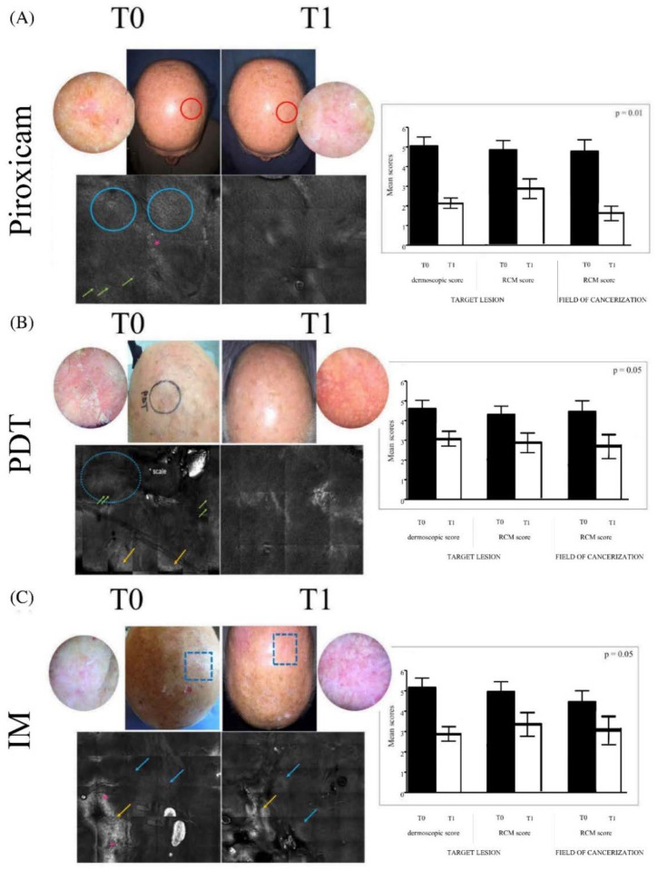
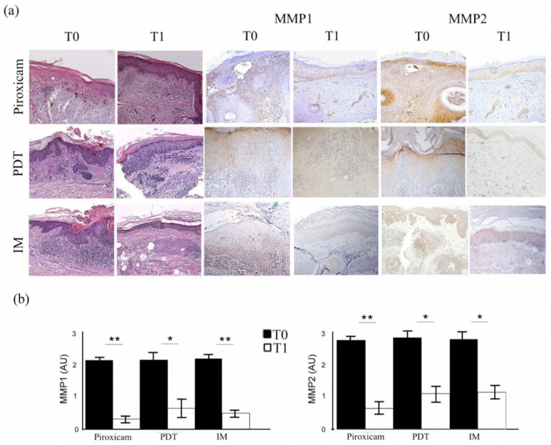
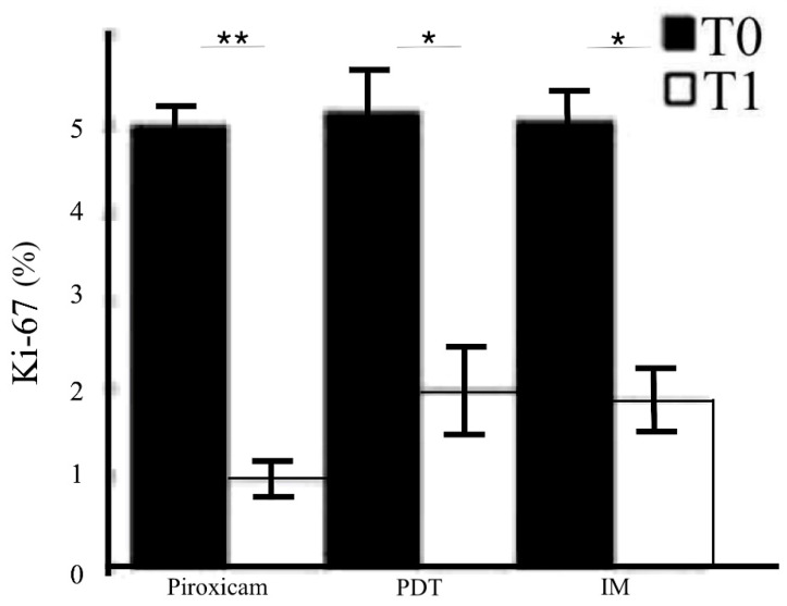
Actinic keratosis is an intraepithelial proliferation of atypical keratinocytes that could progress into invasive squamous cell carcinoma. Most evidence suggests an important role of the dermal matrix metalloproteinases in the progression of atypical skin epithelial lesions. We evaluated the clinical efficacy of three different therapeutic modalities (a medical device containing 0.8% piroxicam cream and 50+ sunscreen, photodynamic therapy, and ingenol mebutate gel) to treat suspicious actinic keratoses, which were biopsied for histopathological examination and then analyzed for the expression of matrix metalloproteinases by immunohistochemistry. Clinical, dermoscopic, and reflectance confocal microscopy evaluations revealed a gradual decrease in all standard scores validated for actinic keratosis assessment at the end of the treatments. From a histopathological point of view, we documented the substantial restoration of normal skin architecture, while the immunohistochemical evaluation of matrix metalloproteinases showed a reduction in expression in the treated skin lesions compared to the baseline. As actinic keratoses are considered the precursors of squamous cell carcinoma, their treatment is crucial to prevent the development of a more aggressive disease. Our study monitored the evolution of actinic keratoses subjected to three different topical therapies, with the value of correlating clinical and histopathological findings. Moreover, as the matrix metalloproteinases are largely recognized factors involved in the pathogenesis and evolution of actinic keratosis to squamous cell carcinoma, the demonstration by immunohistochemistry of a reduction in their expression after the treatments adds new valuable concern to the field.
Keywords: actinic keratosis; immunohistochemistry; ingenol mebutate; metalloproteinases; photodynamic therapy; piroxicam; topical therapy.
Conflict of interest statement
The authors declare no conflict of interest.
Figures
Similar articles
-
Identification of differentially expressed genes in actinic keratosis samples treated with ingenol mebutate gel.PLoS One. 2020 May 15;15(5):e0232146. doi: 10.1371/journal.pone.0232146. eCollection 2020. PLoS One. 2020. PMID: 32413042 Free PMC article.
-
Randomized, double-blind, double-dummy, vehicle-controlled study of ingenol mebutate gel 0.025% and 0.05% for actinic keratosis.J Am Acad Dermatol. 2009 Jun;60(6):934-43. doi: 10.1016/j.jaad.2009.01.008. J Am Acad Dermatol. 2009. PMID: 19467365 Clinical Trial.
-
Treatment of facial actinic keratoses with aminolevulinic acid photodynamic therapy (ALA-PDT) or ingenol mebutate 0.015% gel with and without prior treatment with ALA-PDT.J Drugs Dermatol. 2014 Nov;13(11):1353-6. J Drugs Dermatol. 2014. PMID: 25607702 Clinical Trial.
-
Ingenol mebutate gel 0.015% and 0.05%: in actinic keratosis.Drugs. 2012 Dec 24;72(18):2397-405. doi: 10.2165/11470090-000000000-00000. Drugs. 2012. PMID: 23231025 Review.
-
Pharmacokinetic and pharmacodynamic evaluation of ingenol mebutate for the treatment of actinic keratosis.Expert Opin Drug Metab Toxicol. 2018 Sep;14(9):911-918. doi: 10.1080/17425255.2018.1508449. Epub 2018 Aug 9. Expert Opin Drug Metab Toxicol. 2018. PMID: 30074409 Review.
Cited by
-
A Panel of Potential Serum Markers Related to Angiogenesis, Antioxidant Defense and Hypoxia for Differentiating Cutaneous Squamous Cell Carcinomas from Actinic Keratoses.J Pers Med. 2024 Jan 17;14(1):103. doi: 10.3390/jpm14010103. J Pers Med. 2024. PMID: 38248804 Free PMC article.
-
Preliminary Evidence of Efficacy, Safety, and Treatment Satisfaction with Tirbanibulin 1% Ointment: A Clinical Perspective on Actinic Keratoses.Pharmaceuticals (Basel). 2023 Dec 4;16(12):1686. doi: 10.3390/ph16121686. Pharmaceuticals (Basel). 2023. PMID: 38139813 Free PMC article.
-
Field Cancerization Therapies for the Management of Actinic Keratosis: An Updated Review.Am J Clin Dermatol. 2024 May;25(3):391-405. doi: 10.1007/s40257-023-00839-8. Epub 2024 Feb 13. Am J Clin Dermatol. 2024. PMID: 38351246 Review.
-
Transcriptomic Study on Human Skin Samples: Identification of Two Subclasses of Actinic Keratoses.Int J Mol Sci. 2023 Mar 21;24(6):5937. doi: 10.3390/ijms24065937. Int J Mol Sci. 2023. PMID: 36983009 Free PMC article.
-
Targeting Biomarkers of Proliferation and Inflammation (Ki67, p53, and COX-2) in Actinic Keratoses with Photodynamic Therapy.Biomedicines. 2025 Jun 17;13(6):1487. doi: 10.3390/biomedicines13061487. Biomedicines. 2025. PMID: 40564206 Free PMC article.
References
-
- Röwert-Huber J., Patel M.J., Forschner T., Ulrich C., Eberle J., Kerl H., Sterry W., Stockfleth E. Actinic keratosis is an early in situ squamous cell carcinoma: A proposal for reclassification. Br. J. Dermatol. 2007;156((Suppl. 3)):8–12. doi: 10.1111/j.1365-2133.2007.07860.x. Erratum in Br. J. Dermatol. 2007, 157, 431. - DOI - PubMed
MeSH terms
Substances
LinkOut - more resources
Full Text Sources
Medical
Research Materials
