

Gadolinium Accumulation and Toxicity on In Vitro Grown Stevia rebaudiana: A Case-Study on Gadobutrol
- PMID: 36232670
- PMCID: PMC9569896
- DOI: 10.3390/ijms231911368
Gadolinium Accumulation and Toxicity on In Vitro Grown Stevia rebaudiana: A Case-Study on Gadobutrol
Abstract
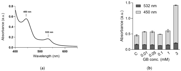
Gadolinium-based contrast agents are molecular complexes which are extensively used for diagnostic purposes. Apart from their tremendous contribution to disease diagnostics, there are several issues related to their use. They are extremely stable complexes and potential contaminants of surface and ground waters, an issue which is documented worldwide. The irrigation of fields with contaminated surface waters or their fertilization with sludge from wastewater treatment plants can lead to the introduction of Gd into the human food supply chain. Thus, this study focused on the potential toxicity of Gd on plants. For this purpose, we have studied the molecular effects of gadobutrol (a well-known MRI contrast agent) exposure on in vitro-grown Stevia rebaudiana. The effects of gadobutrol on plant morphology, on relevant plant metabolites such as chlorophylls, carotenoids, ascorbic acids (HPLC), minerals (ICP-OES), and on the generation of free radical species (MDA assay and EPR) were assessed. Exposures of 0.01, 0.05, 0.1, 1, and 3 mM gadobutrol were used. We found a correlation between the gadobutrol dose and the plant growth and concentration of metabolites. Above the 0.1. mM dose of gadobutrol, the toxic effects of Gd+3 ions became significant.
Keywords: accumulation; carotenoids; chlorophylls; free radicals; gadobutrol; growth; toxicity.
Conflict of interest statement
The authors declare no conflict of interest.
Figures


References
-
- Errante Y., Cirimele V., Mallio C.A., Di Lazzaro V., Zobel B.B., Quattrocchi C.C. Progressive increase of T1 signal intensity of the Dentate nucleus on unenhanced magnetic resonance images is associated with cumulative doses of intravenously administered gadodiamide in patients with normal renal function, suggesting dechelation. Investig. Radiol. 2014;49:685–690. doi: 10.1097/RLI.0000000000000072. - DOI - PubMed
-
- Kanda T., Ishii K., Kawaguchi H., Kitajima K., Takenaka D. High Signal Intensity in the Dentate Nucleus and Globus Pallidus on Unenhanced T1-weighted MR Images: Relationship with Increasing Cumulative Dose of a Gadolinium-based Contrast Material. Radiology. 2014;270:834–841. doi: 10.1148/radiol.13131669. - DOI - PubMed
MeSH terms
Substances
Grants and funding
LinkOut - more resources
Full Text Sources
