. 2022 Sep 28;23(19):11438.
doi: 10.3390/ijms231911438.

Pancreatic Cancer Cells Induce MicroRNA Deregulation in Platelets

Affiliations

Pancreatic Cancer Cells Induce MicroRNA Deregulation in Platelets

Jorge Yassen Díaz-Blancas et al. Int J Mol Sci. .

Abstract

Pancreatic cancer is a pathology with a high mortality rate since it is detected at advanced stages, so the search for early-stage diagnostic biomarkers is essential. Liquid biopsies are currently being explored for this purpose and educated platelets are a good candidate, since they are known to present a bidirectional interaction with tumor cells. In this work, we analyzed the effects of platelets on cancer cells' viability, as determined by MTT, migration using transwell assays, clonogenicity in soft agar and stemness by dilution assays and stem markers' expression. We found that the co-culture of platelets and pancreatic cancer cells increased the proliferation and migration capacity of BXCP3 cells, augmented clonogenicity and induced higher levels of Nanog, Sox2 and Oct4 expression. As platelets can provide horizontal transfer of microRNAs, we also determined the differential expression of miRNAs in platelets obtained from a small cohort of pancreatic cancer patients and healthy subjects. We found clear differences in the expression of several miRNAs between platelets of patients with cancer healthy subjects. Moreover, when we analyzed microRNAs from the platelets of the pancreatic juice and blood derived from each of the cancer patients, interestingly we find differences between the blood- and pancreatic juice-derived platelets suggesting the presence of different subpopulations of platelets in cancer patients, which warrant further analysis.

Keywords: miRNAs; pancreas cancer; tumor-educated platelets.


Conflict of interest statement

The authors declare no conflict of interest. The funders had no role in the design of the study; in the collection, analyses, or interpretation of data; in the writing of the manuscript; or in the decision to publish the results.

Figures
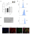
Figure 1
Figure 1
In vitro platelet production by different inductors. (A) MEG−01-derived platelets were counted after 72 h of treatment with: VPA (acid valproic), TPO (thrombopoietin) and a combination of both. Platelets were manually counted in a Neubauer chamber. The asterisks indicate a p < 0.005. Inset: Left: Photograph of CaCl2-induced clot formed by platelets co-cultured with BxPC−3 cancer cells (left panel, 1) or MEG−01-derived platelets; (ns: non-stained platelets) (B) Representative flow cytometry histogram of CD41-labelled platelets. Top panel: control (ns: non-stained platelets) isolated from healthy donors; middle panel: platelets produced by MEG-01 differentiation and lower panel: platelets isolated from healthy donors; (C) Photograph of platelets co-cultured with BxPC−3 cancer cells; (D) Immunofluorescence of BxPC−3 cells in co-culture with platelets. CD41 was stained with AlexaFlour488 (green), membranes were stained with FM 4-64 dye (red) and their nuclei were stained with DAPI (blue). *** p < 0.001, **** p < 0.0001.
Figure 2
Figure 2
Effects of the platelets on cancer cells. (A) Viability curves of BxPC−3 cells exposed to platelets for 24, 48, and 72 h using the MTT assay; (ns: non-stained platelets) (B) Clonogenicity assay; 9 serial dilutions of cells were made, cultured for two weeks, and stained with crystal violet. Both groups were observed and quantified at 3 time points: 24 h, 48 h and 72 h; (C) Representative clonogenicity assays of unexposed cells (left) or cells exposed to platelets (right) after 7 days of culture on a double layer of semi-solid agar [40]; (D) Migration of BxPC−3 cells exposed and not exposed to platelets for 48 hrs; (E) Example image of a migration assay of BxPC−3 cells exposed to platelets. ns: not significant, * p < 0.05, ** p < 0.01, **** p < 0.0001.
Figure 3
Figure 3
Cancer cells exposed to platelets present stem-like characteristics. (A) Clonogenicity assay using 9 serial dilutions of ΒxPC−3 cells exposed to platelets; (B) and (C) Calculation of the proportion of stem cells present in the BxPC−3 culture by the ELDA method with (red) or without (black) platelets. The presence of platelets increases the ratio of stem cells of BxPC−3 cells about 10 times more than in control cells; (D) qPCR quantification of 3 genes directly associated with the self-renewal and pluripotent state characteristic of cancer stem cells. * p < 0.05, ** p < 0.01.
Figure 4
Figure 4
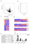
Differentially expressed miRNAs in platelets derived from pancreatic cancer patients (Tumor Educated Platelets). A total of 4 blood samples and 4 pancreatic juice samples were taken from 4 different patients of pancreatic cancer. These samples were paired with 4 blood samples from healthy donors. (A) Volcano plot that shows statistical (p value) versus magnitude of change (fold change) of differentially expressed platelet transcriptome of patients with tumor cancer and of people without cancer; (B) Principal component analysis of platelets’ transcriptome of patients with tumor cancer and of people without cancer; (C) Unsupervised hierarchical clustering of all samples. Two groups were found that discriminates between pancreatic cancer and control individuals: (D) Clustering heatmap of pancreatic drainage samples vs. control blood. Top panel: Two groups were found by k-means clustering algorithm. Heatmap of blood samples cancer vs. control by k-means clustering middle panel. Heat map of platelets from blood (Bp) or platelets of pancreatic juice (Pjp) cancer; (E) Result of the machine-learning signal-noise analysis between cancer samples, in this case we selected the top miRNAs to 8 candidates with an optimal p-value; (F) Top 4 differentially expressed miRNAs quantification by qPCR from platelets trained with direct contact with the BxPC-3 cells against control platelets (* p < 0.05, ** p < 0.01, *** p < 0.001, **** p < 0.0001).

References

    1. Mai S., Inkielewicz-Stepniak I. Pancreatic Cancer and Platelets Crosstalk: A Potential Biomarker and Target. Front. Cell Dev. Biol. 2021;9:749689. doi: 10.3389/fcell.2021.749689. - DOI - PMC - PubMed
    1. Bray F., Ferlay J., Soerjomataram I., Siegel R.L., Torre L.A., Jemal A. Global cancer statistics 2018: GLOBOCAN estimates of incidence and mortality worldwide for 36 cancers in 185 countries. CA Cancer J. Clin. 2018;68:394–424. doi: 10.3322/caac.21492. - DOI - PubMed
    1. Zou X., Wei J., Huang Z., Zhou X., Lu Z., Zhu W., Miao Y. Identification of a six-miRNA panel in serum benefiting pancreatic cancer diagnosis. Cancer Med. 2019;8:2810–2822. doi: 10.1002/cam4.2145. - DOI - PMC - PubMed
    1. Lewis A.R., Valle J.W., McNamara M.G. Pancreatic cancer: Are “liquid biopsies” ready for prime-time? World J. Gastroenterol. 2016;22:7175–7185. doi: 10.3748/wjg.v22.i32.7175. - DOI - PMC - PubMed
    1. Moutinho-Ribeiro P., Macedo G., Melo S.A. Pancreatic Cancer Diagnosis and Management: Has the Time Come to Prick the Bubble? Front. Endocrinol. 2018;9:779. doi: 10.3389/fendo.2018.00779. - DOI - PMC - PubMed