. 2022 Sep 29;23(19):11505.
doi: 10.3390/ijms231911505.

Characterization of New Defensin Antimicrobial Peptides and Their Expression in Bed Bugs in Response to Bacterial Ingestion and Injection

Affiliations

Characterization of New Defensin Antimicrobial Peptides and Their Expression in Bed Bugs in Response to Bacterial Ingestion and Injection

Sanam Meraj et al. Int J Mol Sci. .

Abstract

Common bed bugs, Cimex lectularius, can carry, but do not transmit, pathogens to the vertebrate hosts on which they feed. Some components of the innate immune system of bed bugs, such as antimicrobial peptides (AMPs), eliminate the pathogens. Here, we determined the molecular characteristics, structural properties, and phylogenetic relatedness of two new defensins (CL-defensin1 (XP_024085718.1), CL-defensin2 (XP_014240919.1)), and two new defensin isoforms (CL-defensin3a (XP_014240918.1), CL-defensin3b (XP_024083729.1)). The complete amino acid sequences of CL-defensin1, CL-defensin2, CL-defensin3a, and CL-defensin3b are strongly conserved, with only minor differences in their signal and pro-peptide regions. We used a combination of comparative transcriptomics and real-time quantitative PCR to evaluate the expression of these defensins in the midguts and the rest of the body of insects that had been injected with bacteria or had ingested blood containing the Gram-positive (Gr+) bacterium Bacillus subtilis and the Gram-negative (Gr-) bacterium Escherichia coli. We demonstrate, for the first time, sex-specific and immunization mode-specific upregulation of bed bug defensins in response to injection or ingestion of Gr+ or Gr- bacteria. Understanding the components, such as these defensins, of the bed bugs' innate immune systems in response to pathogens may help unravel why bed bugs do not transmit pathogens to vertebrates.

Keywords: Toll and IMD pathways; bed bugs; defensin antimicrobial peptides; hematophagy; humoral innate immunity; immunization; pathogens; vectors.


Conflict of interest statement

The authors declare no conflict of interest. The funders had no role in the design of the study, in the collection, analyses, or interpretation of data, the writing of the manuscript, or in the decision to publish the results.

Figures
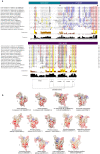
Figure 1
Figure 1
(a) Comparison of structural features of defensins in bed bugs, Cimex lectularius, and other select arthropods. Defensins were aligned using both MUSCLE (multiple sequence comparison by log-expectation; https://www.ebi.ac.uk/Tools/msa/muscle/, accessed on 26 January 2022) and multiple sequence alignment (MSA). Conserved cysteines are yellow-highlighted, positively charged side groups (basic residues; lysine (K), arginine (R), histidine (H)) are red-highlighted, and negatively charged side groups (acidic residues; glutamate (E), aspartate (D)) are blue-highlighted. The predicted signal-peptide, pro-peptide, and mature peptide regions are indicated in boxes above MSA and the cleavage sites between these regions are indicated by arrows. Conserved cysteines and glycines are yellow- and grey-highlighted, respectively. Conserved structural features, including the n-loop, α γ-core, m-loop closer to the numbers, β1, c-loop, β2, and the disulfide bridges, are all presented at the bottom of the figure. The RXXR mature peptide cleavage site motif is outlined by the red rectangle. (b) Comparison of predicted 3D structures of defensins from bed bugs and other select insects (using the protein structure homology-modelling server SWISS-MODEL), and the effect of amino acid substitution on predicted surface charge. Amino acids with basic residues (K, R, H) and acidic residues (E, D) are red- and blue-highlighted, respectively. The net charge of each defensin is represented next to its structure. Three of the bed bug defensins have a charge (5, 7) comparable to the charge (6, 7) of kissing bug, Rhodnius prolixus, defensins. Bed bug and kissing bug defensins, unlike defensins from other hematophagous arthropods and the vinegar fly Drosophila melanogaster have a stronger positive surface charge that interacts with the negative charge on microbial membranes, leading to their depolarization, perforation, and death [47]. The tertiary structures were predicted based on the template 1ica.1.A (Protein Data Bank ID)—3D structure of defensin MGD-1 from blow fly larvae, Phormia terranovae [48]. We used 1ica.1.A as a template because (i) it was highly ranked for all bed bug defensins, and (ii) the redefined 3-dimensional model of defensin A is derived from structural analyses using extensive two-dimensional nuclear magnetic resonance spectroscopy (786 inter-proton nuclear Overhauser effects) [48]. Sequence similarity between 1ica.1.A and the selected defensins was above 65% in all cases. The structures presented were derived from peptides in aqueous solution.
Figure 2
Figure 2
Phylogeny of the mature and active region of defensins identified in bed bugs, Cimex lectularius, and other select arthropods. The defensins isolated from bed bugs are most closely aligned with those from another hemipteran, the kissing bug Rhodnius prolixus. Defensin sequences were aligned with MUSCLE (https://www.ebi.ac.uk/Tools/msa/muscle/, accessed on 26 January 2022), and the alignments were used to build phylogenetic trees using iqtree-2.0-rc2 with substitution models BLOSUM62 and PMB. The tree was finalized using iTOL v6 (https://itol.embl.de/, accessed on 26 January 2022, Heidberg, Germany). Branch lengths are represented on top of each branch. Phylogenetic testing included 10,000 replicates of Ultrafast bootstrap (UFBoot) represented on each branch to provide support for tree branches. The defensin from the tick Haemaphysalis longicornis (ATN39847.1) was used as the outgroup.
Figure 3
Figure 3
Time-dependent expression of CL-defensin1 mRNA (LOC106661793) in bed bugs after bacterial injection. Samples of midgut and RoB (rest of body containing bodies minus heads and midgut tissues) were collected from bed bugs 12 and 24 h after they were injected with a mixture of Gram-positive (Bacillus subtilis ATCC 6633) and Gram-negative (Escherichia coli K12/D31) bacteria. (a) Levels of defensin mRNA in the midgut 12 h and 24 h after bacterial injection. (b) Levels of defensin mRNA in the RoB 12 h and 24 h after bacterial injection. (c) Comparison of the effect of time on the defensin mRNA expression in the midgut and RoB 12 h and 24 h after bacterial injection. The relative expression of defensin was evaluated using the ΔΔCT method [49,50]. The expression level from PBS-control-injected samples was used as the second calibrator and set arbitrarily at 1 (subpanels a and b), and data at the 12-h time point (white bars) were used as the second calibrator in panel c. In panel c, the effect of time (12 h or 24 h) on defensin expression was compared using the formula 2ΔCt 24 h bacterial inj/2ΔCt 12 h bacterial inj. The data representing 12 h after bacterial injection were arbitrarily set to 1. The fold-change of defensin expression 24 h after bacterial injection is shown as grey bars. Bars represent the mean transcript levels ± 95% CI. Means were compared using the unpaired Student’s t-test (* p < 0.05, ** p < 0.01, **** p < 0.0001).
Figure 4
Figure 4
Comparative transcriptome (RNAseq) analyses of CL-defensin1, CL-defensin2, and CL-defensin 3a and 3b expression in bed bugs after ingestion of bacteria-infected blood. Comparative analyses of gene expression in the transcriptome study in bed bug midguts and RoB tissues (rest of body containing bodies minus heads and midgut tissues) after ingestion of sterile blood or blood infected with the Gram-positive bacterium Bacillus subtilis ATCC 6633 or the Gram-negative bacterium Escherichia coli K12/D31. The Wald test was used to generate p-values and Log2 fold changes. An asterisk indicates statistically significant changes in gene expression levels (adjusted p-values < 0.05).
Figure 5
Figure 5
Changes in expression levels of CL-defensin1 in the midguts and RoB (rest of body containing bodies minus heads and midgut tissues) of male (a,b) and female (c,d) bed bugs 12 h after intrathoracic injection or ingestion of Gram-negative (Escherichia coli K12/D31) or Gram-positive (Bacillus subtilis ATCC 6633) bacteria. White bars represent data obtained from control bugs that were injected with phosphate buffer saline (PBS) or that ingested sterile blood. The relative expression of CL-defensin1 (LOC106661793) was evaluated using the ΔΔCT method [49,50]. Bars represent the mean transcript levels ± 95% CI. Means were compared using the unpaired Student’s t-test (* p < 0.05, *** p < 0.001, **** p < 0.0001).
Figure 6
Figure 6
Effect of bed bug sex on changes in expression levels of CL-defensin1 12 h after intrathoracic injection or ingestion of the Gram-positive bacterium Bacillus subtilis (ATCC 6633) or the Gram-negative bacterium Escherichia coli (K12/D31) in the midgut (a) and RoB (rest of body containing bodies minus heads and midgut tissues) (b). The relative expression of defensin was evaluated using the ΔΔCT method [49,50]. Data from males were used as the second calibrator and were arbitrarily set to 1; the fold-changes of defensin expression in females are shown as grey bars. Bars represent the mean transcript levels ± 95% CI. Means were compared using the unpaired Student’s t-test (** p < 0.01, *** p < 0.001, **** p < 0.0001).
Figure 7
Figure 7
Comparison of the mode of infection, bacterial ingestion, or injection, on changes in expression levels of CL-defensin1 in bed bugs. Samples of midgut and RoB (rest of body containing bodies minus heads and midgut tissues) were collected from male (a) and female (b) bed bugs 12 h after the injection of bacteria (Gram-positive Bacillus subtilis ATCC 6633 or Gram-negative bacteria Escherichia coli K12/D31), or 12 h after ingestion of blood infected with E. coli or B. subtilis. The relative expression of defensin was evaluated using the ΔΔCT method [49,50]. The effect of bacterial ingestion versus bacterial injection was compared using the formula 2ΔCt injected/2ΔCt ingested. The ingestion-sample data (white bars) representing the calibrator were arbitrarily set to 1, and fold-changes in defensin expression in bacterial injection sample data are shown as grey bars. Bars represent the mean transcript levels ± 95% CI. Means were compared using the unpaired Student’s t-test (** p < 0.01, **** p < 0.0001).
Figure 8
Figure 8
Effect of blood ingestion by male bed bugs on pH changes in their midgut and RoB (rest of body containing bodies minus heads and midgut tissues). Midgut and RoB tissue were dissected from >28-day-starved bed bugs that ingested blood (“fed”) or did not (“unfed”). Data were collected 24 h after blood ingestion. The pH was measured using the LAQUAtwin pH 22 pH meter (Horiba, Kyoto, Japan). At least five biological replicates were used per treatment or control group and five insects were pooled for each group. Data were analyzed by one-way ANOVA and Brown–Forsythe test (*** p < 0.001, **** p < 0.0001).

Similar articles

Cited by

References

    1. Doggett S.L., Dwyer D.E., Penas P.F., Russell R.C. Bed bugs: Clinical relevance and control options. Clin. Microbiol. Rev. 2012;25:164–192. doi: 10.1128/CMR.05015-11. - DOI - PMC - PubMed
    1. Parola P., Izri A. Bedbugs. N. Engl. J. Med. 2020;382:2230–2237. doi: 10.1056/NEJMcp1905840. - DOI - PubMed
    1. Balster C.B. Master’s Thesis. Wright State University; Dayton, OH, USA: 2011. The Non-Infectious Disease Implications of Bed Bug Infestations.
    1. Burton G.J. Bedbugs in relation to transmission of human diseases. Review of the literature. Public Health Rep. 1963;78:513–524. doi: 10.2307/4591852. - DOI - PMC - PubMed
    1. Delaunay P., Blanc V., Del Giudice P., Levy-Bencheton A., Chosidow O., Marty P., Brouqui P. Bedbugs and infectious diseases. Clin. Infect. Dis. 2011;52:200–210. doi: 10.1093/cid/ciq102. - DOI - PMC - PubMed