. 2022 Sep 29;23(19):11510.
doi: 10.3390/ijms231911510.

Therapeutic Potential of Mesenchymal Stem Cell-Secreted Factors on Delay in Corneal Wound Healing by Nitrogen Mustard

Affiliations

Therapeutic Potential of Mesenchymal Stem Cell-Secreted Factors on Delay in Corneal Wound Healing by Nitrogen Mustard

Seungwon An et al. Int J Mol Sci. .

Abstract

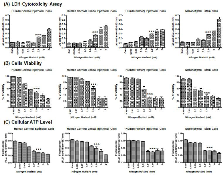
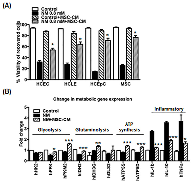
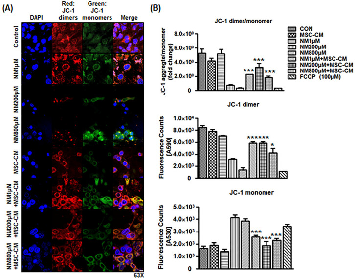
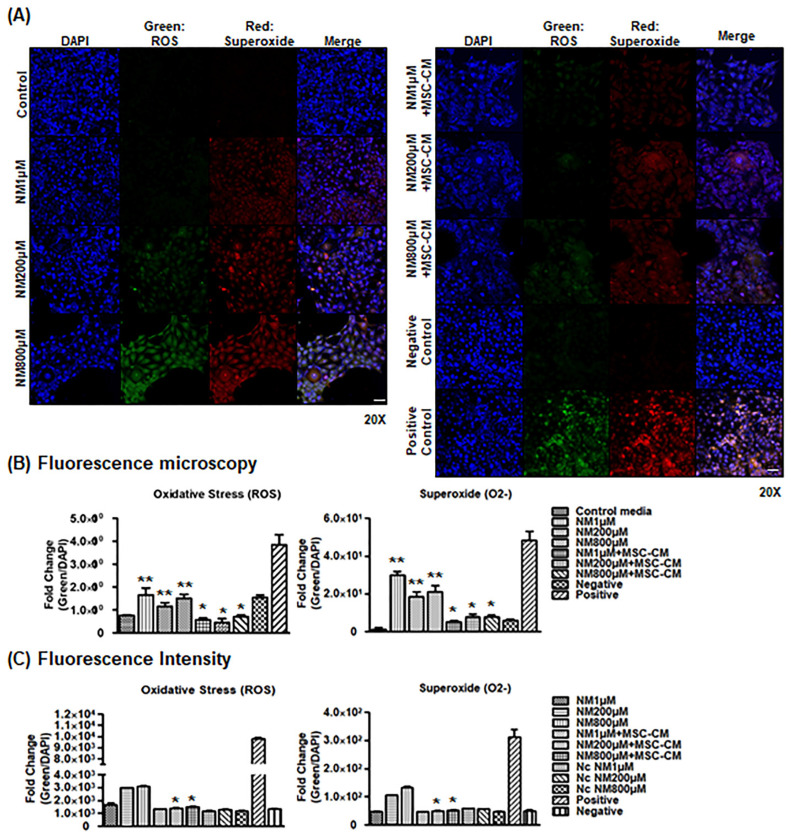
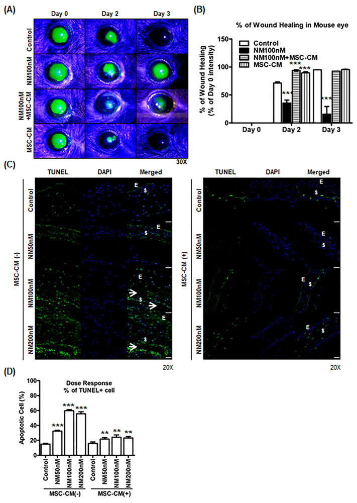
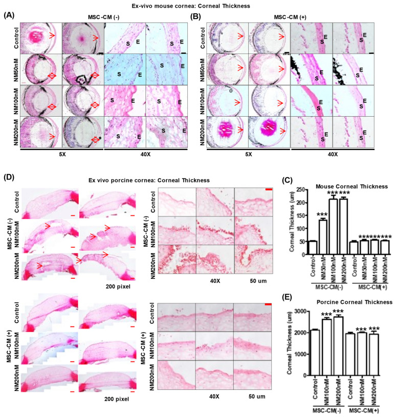
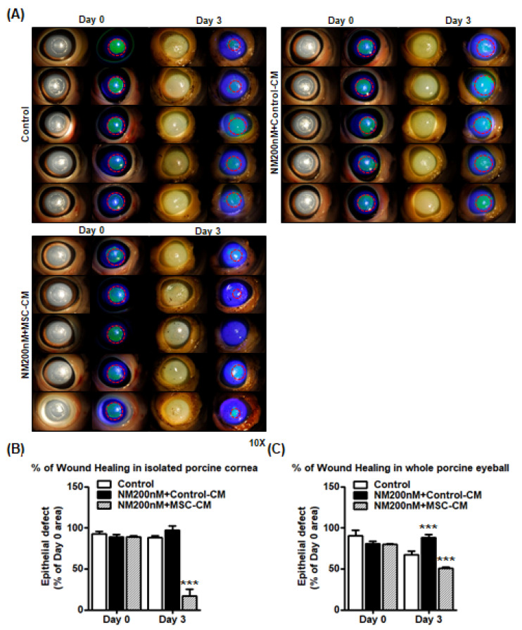
Ocular surface exposure to nitrogen mustard (NM) leads to severe ocular toxicity which includes the separation of epithelial and stromal layers, loss of endothelial cells, cell death, and severe loss of tissue function. No definitive treatment for mustard gas-induced ocular surface disorders is currently available. The research was conducted to investigate the therapeutic potential of mesenchymal stem cell-conditioned media (MSC-CM) in NM-induced corneal wounds. NM was added to different types of corneal cells, the ocular surface of porcine, and the ocular surface of mice, followed by MSC-CM treatment. NM significantly induced apoptotic cell death, cellular ROS (Reactive oxygen species), and reduced cell viability, metabolic gene expression, and mitochondrial function, and, in turn, delayed wound healing. The application of MSC-CM post NM exposure partially restored mitochondrial function and decreased intracellular ROS generation which promoted cell survival. MSC-CM therapy enhanced wound healing process. MSC-CM inhibited NM-induced apoptotic cell death in murine and porcine corneal tissue. The application of MSC-CM following a chemical insult led to significant improvements in the preservation of corneal structure and wound healing. In vitro, ex vivo, and in vivo results suggest that MSC-CM can potentially provide targeted therapy for the treatment of chemical eye injuries, including mustard gas keratopathy (MGK) which presents with significant loss of vision alongside numerous corneal pathologies.

Keywords: apoptotic cell death; cellular ROS; corneal injury; delayed wound healing; mesenchymal stem cell-conditioned media; nitrogen mustard.


Conflict of interest statement

The authors have no conflict of interest to disclose.

Figures

Figure 1
Figure 1
Effects of nitrogen mustard exposure on cell cytotoxicity, cell viability, and ATP level in culture cells. (A) Graph showing cytotoxicity for various doses of nitrogen mustard (NM) in culture cells as measured by LDH assay (n = 6/group). (B) Graph showing cell viability for various doses of NM in culture cells as measured by LDH assay (n = 6/group). (C) Graph showing ATP levels for various doses of NM in culture cells as measured by LDH assay (n = 6/group). *** p < 0.001 vs. untreated control or 0.001 mM NM treated group.
Figure 2
Figure 2
Mesenchymal Stromal Cell (MSCs)-conditioned media recovers immediate cytotoxic effects in the corneal epithelial cells following exposure to nitrogen. (A) Cell viability of recovered cells is measured by trypan blue staining with control, NM 0.8 mM, control media + MSC-CM, and NM 0.8 mM + MSC-CM. *** p < 0.001 vs. NM 0.8 mM. (B) Graph showing the mRNA levels of genes for glycolysis, glutaminolysis, ATP synthesis, ATP synthesis, and inflammatory genes in control, NM 0.8 mM (2 h), and NM 0.8 mM (2 h) + MSC-CM (24 h) by performing qPCR. HCEC cells are used for NM treatment. * p < 0.05, ** p < 0.01, *** p < 0.001 vs. NM treated group.
Figure 3
Figure 3
MSC-CM attenuates NM-exposed mitochondrial membrane potential change. (A) Representative brightfield images after NM exposure or NM with MSC-CM incubation. Representative fluorescence microscopy images of JC-1 dimers (red) and JC-1 monomers (green) after various doses of NM with or without MSC-CM. The merged panel presents co-localization between red dimer and green monomer fluorescence. Scale bar, 10 μm (B) Fluorescence intensity of mitochondria membrane potentials (JC-1 dimer, JC-1 monomers, and JC-1 dimer/monomer) by fluorimetry. FCCP 100 μM was used as depolarization negative control. * p < 0.05, *** p < 0.001 vs. NM treated group.
Figure 4
Figure 4
MSC-CM decreases cellular ROS generation. (A) Representative confocal immunofluorescence microscopy images of ROS (green) and Superoxide (red) after various doses of NM with or without MSC-CM. Scale bar, 50 μm (B) Relative fold changes from Figure 4A. ** p < 0.01 vs. Control media, * p < 0.05 vs. NM treated group. (C) Representative ROS/Superoxide assay showing the effect of MSC-CM on NM exposure in cell culture. Fluorescence intensity after NM (1, 200, and 800 uM) and NM with MSC-CM incubation for 2 h. Positive control (ROS Inducer: Pyocyanin). Negative Control (ROS Inhibitor: N-acetyl-L-cysteine). Negative Control (solvent). * p < 0.05 vs. NM treated group.
Figure 5
Figure 5
Wound healing effect of MSC-CM on NM injury on mouse corneas. (A) Representative images of mouse corneas (n = 4/group) showing fluorescein staining after epithelial scratch and topical application of 10 μL of NM 100 nM on the center of cornea for 30 min. After 30 min, corneas were washed with 1 mL of 1X PBS, and corneas were applied with MSC-CM twice a day for 3 days. (B) Relative fold changes of comparing epithelial defect area between groups at Day 0 to Day 3 (n = 4/group) from Figure 5A. *** p < 0.001 vs. untreated control (Day 2) or untreated control (Day 3). (C) Ex vivo mouse eye culture model: For apoptotic cell death, mouse eyes were cultured in various doses of NM (50, 100, and 200 nM) in KSFM media for 2 h and washed two times with 1X PBS. After washing, NM-exposed mouse eyes were replaced with (B) or without (A) MSC-CM for 2 days and imaged using 20× magnification im-immunofluorescence by TUNEL assay. TUNEL (Green), DAPI (Blue). Scale bar, 50 μm (D) Graphs showing apoptotic cell death on mouse cornea area from Figure 5C. *** p < 0.001 vs. untreated control (MSC-CM-), ** p < 0.01 vs. NM treated group (MSC-CM-).
Figure 6
Figure 6
MSC-CM reverses NM-induced corneal epithelial damage in ex vivo mouse eye culture model. (A,B) Ex vivo mouse eye culture model: Representative images of H&E staining showing histology of mouse corneas after various doses of NM (50, 100, and 200 nM) in KSFM media for 2 h and washing twice with 1× PBS. After washing, NM-exposed mouse eyes were replaced with (B) or without (A) MSC-CM for 2 days and imaged using 5× and 40× magnification microscope. S: Stroma, E: Epithelium. (C) Graphs showing corneal thickness on mouse cornea area from Figure 6A,B. *** p < 0.001 vs. untreated control or NM treated group (MSC-CM-). (D) Ex vivo porcine eye culture model: porcine eyes were cultured in various doses of NM (50, 100, and 200 nM) in KSFM media 2 h and washed two times with 1X PBS. After washing, NM-exposed porcine eyes were replaced with or without MSC-CM for 3 days and imaged using 40× magnification microscope. (E) Porcine corneal thickness was measured from Figure 6D. *** p < 0.001 vs. untreated control or NM treated group (MSC-CM-).
Figure 7
Figure 7
Wound healing effects of MSC-CM on NM injury in ex vivo porcine cornea culture model. (A) Ex vivo porcine cornea culture model for wound healing study: corneas were dissected from whole porcine eyes and a 6.5 mm wound was created on the porcine corneas. The 6.5 mm-wounded porcine corneas were cultured in various doses of NM 200 nM in KSFM media for 2 h and washed two times with 1X PBS. After washing, wounded porcine corneas were incubated with control (1:1 ratio of MEMa and 10% FBS/DMEM), NM 100 nM (in 1:1 ratio of time control media and 10% FBS/DMEM), and NM 200 nM (in 1:1 ratio of MSC-CM and 10% FBS/DMEM) for 3 days. A slit lamp was used to observe corneal staining (10× magnification) on Day 0 and Day 3. (B) Graph comparing epithelial defect area between groups (Control, NM 200 nM, and NM 200 nM + MSC-CM) at Day 0 to Day 3 (n = 5/group). *** p < 0.001 vs. NM 200 nM + Control CM (Day 3), (C) Graph comparing epithelial defect area between groups (Control, NM 200 nM, and NM 200 nM + MSC-CM) at Day 0 to Day 3 (n = 3/group) from Supplementary Figure S3. *** p < 0.001 vs. untreated control (Day 3). These results suggest that MSC-CM have the potential to enhance the healing process. Thus, MSC-CM reversed epithelial thickening and accelerated wound healing after NM injury both in vivo and ex vivo.

Similar articles

Cited by

References

    1. Gordon M.K., Desantis A., Deshmukh M., Lacey C.J., Hahn R.A., Beloni J., Anumolu S.S., Schlager J.J., Gallo M.A., Gerecke D.R., et al. Doxycycline hydrogels as a potential therapy for ocular vesicant injury. J. Ocul. Pharmacol. Ther. 2010;26:407–419. doi: 10.1089/jop.2010.0099. - DOI - PMC - PubMed
    1. Goswami D.G., Kumar D., Tewari-Singh N., Orlicky D.J., Jain A.K., Kant R., Rancourt R.C., Dhar D., Inturi S., Agarwal C., et al. Topical nitrogen mustard exposure causes systemic toxic effects in mice. Exp. Toxicol. Pathol. 2015;67:161–170. doi: 10.1016/j.etp.2014.11.006. - DOI - PMC - PubMed
    1. Goswami D.G., Tewari-Singh N., Dhar D., Kumar D., Agarwal C., Ammar D.A., Kant R., Enzenauer R.W., Petrash J.M., Agarwal R. Nitrogen Mustard-Induced Corneal Injury Involves DNA Damage and Pathways Related to Inflammation, Epithelial-Stromal Separation, and Neovascularization. Cornea. 2016;35:257–266. doi: 10.1097/ICO.0000000000000685. - DOI - PMC - PubMed
    1. Loeber R., Michaelson E., Fang Q., Campbell C., Pegg A.E., Tretyakova N. Cross-linking of the DNA repair protein Omicron6-alkylguanine DNA alkyltransferase to DNA in the presence of antitumor nitrogen mustards. Chem. Res. Toxicol. 2008;21:787–795. doi: 10.1021/tx7004508. - DOI - PMC - PubMed
    1. Jan Y.H., Heck D.E., Laskin D.L., Laskin J.D. DNA damage signaling in the cellular responses to mustard vesicants. Toxicol. Lett. 2020;326:78–82. doi: 10.1016/j.toxlet.2020.03.008. - DOI - PMC - PubMed