. 2022 Sep 29;23(19):11535.
doi: 10.3390/ijms231911535.

Spiropyran/Merocyanine Amphiphile in Various Solvents: A Joint Experimental-Theoretical Approach to Photophysical Properties and Self-Assembly

Affiliations

Spiropyran/Merocyanine Amphiphile in Various Solvents: A Joint Experimental-Theoretical Approach to Photophysical Properties and Self-Assembly

Vladyslav Savchenko et al. Int J Mol Sci. .

Abstract

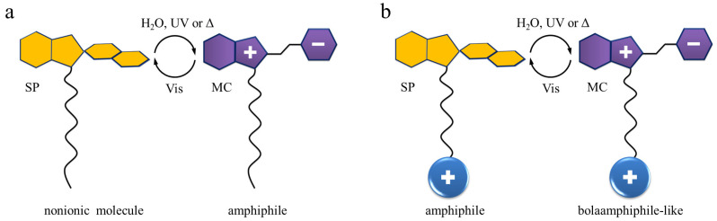
This joint experimental-theoretical work focuses on molecular and photophysical properties of the spiropyran-containing amphiphilic molecule in organic and aqueous solutions. Being dissolved in tested organic solvents, the system demonstrates positive photochromism, i.e., upon UV stimulus the colorless spiropyran form is transformed into colorful merocyanine isomer. However, the aqueous solution of the amphiphile possesses a negative photochromism: the orange-red merocyanine form becomes thermodynamically more stable in water, and both UV and vis stimuli lead to the partial or complete photobleaching of the solution. The explanation of this phenomenon is given on the basis of density functional theory calculations and classical modeling including thermodynamic integration. The simulations reveal that stabilization of merocyanine in water proceeds with the energy of ca. 70 kJ mol-1, and that the Helmholtz free energy of hydration of merocyanine form is 100 kJ mol-1 lower as compared to the behavior of SP isomer in water. The explanation of such a difference lies in the molecular properties of the merocyanine: after ring-opening reaction this molecule transforms into a zwitterionic form, as evidenced by the electrostatic potential plotted around the opened form. The presence of three charged groups on the periphery of a flat conjugated backbone stimulates the self-assembly of merocyanine molecules in water, ending up with the formation of elongated associates with stack-like building blocks, as shown in molecular dynamics simulations of the aqueous solution with the concentration above critical micelle concentration. Our quantitative evaluation of the hydrophilicity switching in spiropyran/merocyanine containing surfactants may prompt the search for new systems, including colloidal and polymeric ones, aiming at remote tuning of their morphology, which could give new promising shapes and patterns for the needs of modern nanotechnology.

Keywords: molecular modeling; negative photochromism; spiropyran/merocyanine isomerization; time-resolved UV-vis measurements.


Conflict of interest statement

The authors declare no conflict of interest.

Figures

Figure 1
Figure 1
Schematic representation of the surfactants and their mutual interconversion under various stimuli: SP/MC is considered as a head (a) or as a part of the tail (b). We underline here different properties of the molecules prior and after isomerization. More details can be found in the main text.
Figure 2
Figure 2
Spiropyran/merocyanine photochromic amphiphilic couple studied in this paper. This surfactant contains a hydrophobic propyl (C3) hydrocarbon chain terminated with a permanently charged quaternary ammonium headgroup and introduced in the ω-position of spiropyran (left) bearing an –NO2 substituent in para-position to the pyran oxygen. It was reported that the electron-acceptor substituents in para-position stabilize the zwitterionic MC form (right) and drastically lower the pKa [11,62].
Figure 3
Figure 3
Snapshots of spiropyran ((a), TMAB-C3-SP) and merocyanine ((b), TMAB-C3-MC) surfactants with Br anions. The structures are as received after geometry optimization in implicit water. Carbon, oxygen, nitrogen, hydrogen and bromine are shown as grey, red, blue, white and burgundy spheres, respectively. The Br anion is in the vicinity of trimethylammonium head, symmetrically centred between three methyl groups of the TMA with the distance N+ – Br of 4.23 ± 0.06 Å on average. The abbreviations TMAB, C3, and SP/MC stand for trimethylammonium bromide, propyl chain, and spiropyran/merocyanine molecular segment, respectively. The multiplicity of the chemical bonds is not shown here, except for both benzene rings and nitro groups. Cartesian coordinates of both isomers are provided in Supplementary Information.
Figure 4
Figure 4
The surfaces of the electrostatic potential around spiropyran (a) and merocyanine (b) cations paired with bromide anion. The surface having a negative sign is colored in blue, the positive one-in red. This picture provides the differences in the V(r) caused by the isomerisation upon light. For this illustration, the following software is utilized: Multiwfn, Version 3.8 [77] and VMD, Version 1.9.3. [78]. The carbon atoms here are colored in cyan, the remaining atoms have colors as noted in Figure 3. The bond order is not shown.
Figure 5
Figure 5
The energy difference ΔEIEFPCMsolvent between the value of a cation in a particular solvent Es and the same in vacuum Ev, as calculated in DFT approach (a) and the Helmholtz solvation free energy ΔFexplicit solvent (b) for cations, as received in thermodynamic integration. The data for chloroform, ethanol, acetonitrile, dimethyl sulfoxide and water are shown in red, olive, black, blue and violet, respectively, for merocyanine (closed vertical columns) and spiropyran (shaded vertical columns). The numbers next to the solvents in panel (a) are the values of ϵ.
Figure 6
Figure 6
The “rainbow” of 0.1 mM spiropyran solution in different solvents under various light stimuli. The “yellow light” means light in the laboratory where the experiments are conducted, which is in our case 570 nm.
Figure 7
Figure 7
Normalized absorption spectra of 0.1 mM SP before irradiation: sample dissolved in appropriate solvent and measured after one hour standing in yellow light lab (a) and normalized absorption spectra of 0.1 mM SP after irradiation (b): samples irradiated with UV light until photostationary state is achieved. The calculated spectra (PBE/6-311G*) of SP (c) and MC (d) forms in different IEFPCM solvents. Computed spectra have been smoothed using Gaussian functions of a half-width at a half-height of 0.333 eV.
Figure 8
Figure 8
Molecular orbitals involved in the electronic transitions of different isomers at absorption bands shown in Figure 7c,d for SP (a) and MC (b). For this illustration, Multiwfn, Version 3.8 [77] has been used.
Figure 9
Figure 9
Normalized absorption spectra of 0.1 mM SP in water. (a,b)—Relaxation in the dark; a sample dissolved in water and placed directly in the spectrometer. (c,d)—The conversion kinetics of the relaxed state (24 h yellow light lab) to UV (365 nm) irradiated photostationary state; the irradiation intensity is 2 mW cm2. (e,f)—The conversion kinetics of the relaxed state (24 h yellow light lab) to blue (455 nm) irradiated photostationary state; the irradiation intensity is 2 mW cm2. The min MC and max MC content is 3 and 100% (panel b). A half-lifetime for complete switching to max MC containing photostationary state is 110 min. For panel d, the max MC is 39% and min MC is 12%. For panel f, these values correspond to 36 and 3%, respectively. These values are obtained using methodology described in our previous publication [93].
Figure 10
Figure 10
The self-assembled structures of the SP (a) and MC (b) as found in MD simulations in water. The aggregates consist of 12 molecules and 12 counterions, which in the case of SP are only partially condensed on the micelle. Less elongated aggregate is observed for spiropyran with the Connolly surface occupied volume of 5201.3 Å3 and the surface area of 4238.0 Å2. More extended micelle is built from MC form. Here, the Connolly surface occupied a volume of 5621.3 Å3 and a surface area of 4066.4 Å2. For MC, the Br ions are predominantly in the vicinity of the TMAB head.
Figure 11
Figure 11
The snapshots of the pairs of SP (a) and MC (b) after geometry optimization. The structure (although we cannot call it the prevailing one, since SP dimers do not have a pattern in common) of the SP dimer given as an example (a). This dimer is stabilized apparently by van der Waals intermolecular contacts (green dashed line). The MC dimer (b) depicted here has a typical mutual arrangement of the molecules. The interactions stabilizing this micellar building block are the ππ interaction schematically shown by a yellow arrow, the van der Waals contacts of alkyls and aryls (green dashed lines), and the electrostatic attractions (crimson dashed lines) related to the pairing of partially negatively charged oxygens of nitro groups with positively charged hydrogens of trimethyl ammonium of a neighboring molecule. Both van der Waals and electrostatic through-space couplings happen on both sides of the stacked dimer. An average length of the ππ distance is 4.11 ± 0.51 Å, of the electrostatic interactions is 2.19 ± 0.07 Å, and the van der Waals contacts are 2.40 ± 0.19 Å in length. Partial charges of O(–NO2) and H(–N(CH3)3) are written in red and gray color, respectively. Visualization is done in Materials Studio 9.0 [96].
Figure 12
Figure 12
The scheme of synthesis of the 1-(3-trimethylammoniopropyl)-3,3-dimethyl-6-nitrospyro(2H-1-benzopyran-2,2-indoline) bromide according to Hammarson et al. [11].

References

    1. Chen S., Costil R., Leung F.K.-C., Feringa B.L. Self-Assembly of Photoresponsive Molecular Amphiphiles in Aqueous Media. Angew. Chem. 2021;133:11708–11731. doi: 10.1002/ange.202007693. - DOI - PMC - PubMed
    1. Volarić J., Szymanski W., Simeth N.A., Feringa B.L. Molecular Photoswitches in Aqueous Environments. Chem. Soc. Rev. 2021;50:12377–12449. doi: 10.1039/D0CS00547A. - DOI - PMC - PubMed
    1. Montagna M., Guskova O. Photosensitive Cationic Azobenzene Surfactants: Thermodynamics of Hydration and the Complex Formation with Poly (methacrylic acid) Langmuir. 2018;34:311–321. doi: 10.1021/acs.langmuir.7b03638. - DOI - PubMed
    1. Hayashita T., Kurosawa T., Miyata T., Tanaka K., Igawa M. Effect of Structural Variation within Cationic Azo-Surfactant upon Photoresponsive Function in Aqueous Solution. Colloid Polym. Sci. 1994;272:1611–1619. doi: 10.1007/BF00664729. - DOI
    1. Santer S. Remote Control of Soft Nano-Objects by Light Using Azobenzene Containing Surfactants. J. Phys. D Appl. Phys. 2017;51:013002. doi: 10.1088/1361-6463/aa95ca. - DOI

LinkOut - more resources