Prognostic and Immunological Role of STK38 across Cancers: Friend or Foe?
- PMID: 36232893
- PMCID: PMC9570386
- DOI: 10.3390/ijms231911590
Prognostic and Immunological Role of STK38 across Cancers: Friend or Foe?
Abstract
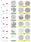
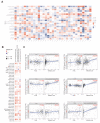
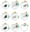
Although STK38 (serine-threonine kinase 38) has been proven to play an important role in cancer initiation and progression based on a series of cell and animal experiments, no systemic assessment of STK38 across human cancers is available. We firstly performed a pan-cancer analysis of STK38 in this study. The expression level of STK38 was significantly different between tumor and normal tissues in 15 types of cancers. Meanwhile, a prognosis analysis showed that a distinct relationship existed between STK38 expression and the clinical prognosis of cancer patients. Furthermore, the expression of STK38 was related to the infiltration of immune cells, such as NK cells, memory CD4+ T cells, mast cells and cancer-associated fibroblasts in a few cancers. There were three immune-associated signaling pathways involved in KEGG analysis of STK38. In general, STK38 shows a significant prognostic value in different cancers and is closely associated with cancer immunity.
Keywords: STK38; immune infiltration; pan-cancer; prognosis; survival.
Conflict of interest statement
The authors declare no conflict of interest.
Figures


Similar articles
-
The Role of Mammalian STK38 in DNA Damage Response and Targeting for Radio-Sensitization.Cancers (Basel). 2023 Mar 30;15(7):2054. doi: 10.3390/cancers15072054. Cancers (Basel). 2023. PMID: 37046714 Free PMC article. Review.
-
The HSP90 inhibitor 17-allylamino-17-demethoxygeldanamycin modulates radiosensitivity by downregulating serine/threonine kinase 38 via Sp1 inhibition.Eur J Cancer. 2013 Nov;49(16):3547-58. doi: 10.1016/j.ejca.2013.06.034. Epub 2013 Jul 22. Eur J Cancer. 2013. PMID: 23886587
-
A Pan-Cancer Analysis of the Immunological and Prognostic Role of BUB1 Mitotic Checkpoint Serine/Threonine Kinase B (BUB1B) in Human Tumors.Clin Lab. 2024 Jan 1;70(1). doi: 10.7754/Clin.Lab.2023.230632. Clin Lab. 2024. PMID: 38213208
-
Grass carp STK38 regulates IFN I expression by decreasing the phosphorylation level of GSK3β.Dev Comp Immunol. 2019 Oct;99:103410. doi: 10.1016/j.dci.2019.103410. Epub 2019 Jun 5. Dev Comp Immunol. 2019. PMID: 31175887
-
The hippo kinase STK38 ensures functionality of XPO1.Cell Cycle. 2020 Nov;19(22):2982-2995. doi: 10.1080/15384101.2020.1826619. Epub 2020 Oct 5. Cell Cycle. 2020. PMID: 33017560 Free PMC article. Review.
Cited by
-
Nanovaccine targeting in colorectal cancer: a multi-dataset analysis of CEA expression, cytokine profiles, and co-expressed genes.Med Pharm Rep. 2025 Jul;98(3):358-370. doi: 10.15386/mpr-2917. Epub 2025 Jul 30. Med Pharm Rep. 2025. PMID: 40786198 Free PMC article.
-
The Role of Mammalian STK38 in DNA Damage Response and Targeting for Radio-Sensitization.Cancers (Basel). 2023 Mar 30;15(7):2054. doi: 10.3390/cancers15072054. Cancers (Basel). 2023. PMID: 37046714 Free PMC article. Review.
-
Methionine restriction and cancer treatment: a systems biology study of yeast to investigate the possible key players.Turk J Biol. 2023 May 23;47(3):208-217. doi: 10.55730/1300-0152.2656. eCollection 2023. Turk J Biol. 2023. PMID: 37529420 Free PMC article.
-
Integrative analysis of the expression profile and prognostic values of SENP gene family in hepatocellular carcinoma.Discov Oncol. 2025 May 13;16(1):752. doi: 10.1007/s12672-025-02598-w. Discov Oncol. 2025. PMID: 40358846 Free PMC article.
-
NDR1 mediates PD-L1 deubiquitination to promote prostate cancer immune escape via USP10.Cell Commun Signal. 2024 Sep 3;22(1):429. doi: 10.1186/s12964-024-01805-5. Cell Commun Signal. 2024. PMID: 39227807 Free PMC article.
References
-
- Yuan S., Norgard R.J., Stanger B.Z. Cellular Plasticity in Cancer. Cancer Discov. 2019;9:837–851. doi: 10.1158/2159-8290.CD-19-0015. - DOI - PMC - PubMed
MeSH terms
Substances
Grants and funding
- No.2019J01016/Natural Science Foundation of Fujian Province of China
- No. 81972373/National Natural Science Foundation of China
- No. 3502Z20194038/Science and Technology Planned Project of Medical and Health of Xiamen City
- No. PM201809170001/Scientific Research Foundation for Advanced Talents of Xiang'an Hospital of Xiamen University
LinkOut - more resources
Full Text Sources
Medical
Molecular Biology Databases
Research Materials

