. 2022 Oct 6;23(19):11857.
doi: 10.3390/ijms231911857.

Ribosomal RACK1 Regulates the Dendritic Arborization by Repressing FMRP Activity

Affiliations

Ribosomal RACK1 Regulates the Dendritic Arborization by Repressing FMRP Activity

Nicla Romano et al. Int J Mol Sci. .

Abstract

FMRP is an RNA-binding protein that represses the translation of specific mRNAs. In neurons, its depletion determines the exaggerated translation of mRNAs leading to dendritic and axonal aberrant development, two peculiar features of Fragile X syndrome patients. However, how FMRP binds to translational machinery to regulate the translation of its mRNA targets is not yet fully understood. Here, we show that FMRP localizes on translational machinery by interacting with the ribosomal binding protein, Receptor for Activated C Kinase 1 (RACK1). The binding of FMRP to RACK1 removes the translational repressive activity of FMRP and promotes the translation of PSD-95 mRNA, one specific target of FMRP. This binding also results in a reduction in the level of FMRP phosphorylation. We also find that the morphological abnormalities induced by Fmr1 siRNA in cortical neurons are rescued by the overexpression of a mutant form of RACK1 that cannot bind ribosomes. Thus, these results provide a new mechanism underlying FMRP activity that contributes to altered development in FXS. Moreover, these data confirm the role of ribosomal RACK1 as a ribosomal scaffold for RNA binding proteins.

Keywords: FMRP; RACK1; neuroblastoma; neuron differentiation; neurons; ribosomes; translation.


Conflict of interest statement

The authors declare no conflict of interest.

Figures

Figure 1
Figure 1
The ribosomal RACK1 regulates dendritic arborization. (A) Immunoblotting for RACK1 and actin on total protein lysate from embryonic mouse hippocampal neurons cultured from 1 DIV to 17 DIV. (Lower) The graph reports the means of three independent experiments of density ratio between RACK1 and actin bands (mean ± S.D.). Student’s T-test was used to calculate p values: * p ≤ 0.05 vs. 1 DIV; # p ≤ 0.01 vs. 7 DIV. B) Immunofluorescence for GFP on hippocampal neurons transfected at 15 DIV with GFP, GFP/RACK1WT-his-myc (WT in the figure), GFP/RACK1DE-his-myc (DE) or RACK1 siRNA (siRNA). Scale 10μm. (C) The graph of Scholl analysis performed on cells in (B). The values were measured as means of the number of neurite intersections measured by Sholl analysis. Data are graphed as mean ± S.D. Student’s T-test was used to calculate p values: * p ≤ 0.01, vs. GFP; # p ≤ 0.05 vs. RWT-his-myc; (D) Graphic of PSD-95, Bassoon density and Pearson’s coefficient of puncta between PSD-95 and Bassoon measured by immunofluorescence experiments. Graph reporting the correlation measured by Pearson coefficient of the puncta signal between PSD-95 and Bassoon in Supplemental Figure S2. All bars represent mean ± SD; n.70 puncta from three independent experiments. One-way ANOVA with Tukey’s Post Hoc test. Data are graphed as mean ± S.E.* p ≤ 0.005, vs. GFP; # p ≤ 0.05, vs. WT.
Figure 2
Figure 2
The dendritic spines and synaptic formation defects of mature Fmr1 siRNA neurons are rescued by the RACK1 mutant up-regulation. (A) GFP immunofluorescence on 11 DIV neurons co-transfected with GFP and Fmr1 siRNA/WT or Fmr1 siRNA/DE. (Lower) In the box is the magnification of neuritis. Scale 20μm upper and 5μm lower. (B) Graphic of PSD-95, Bassoon density and Pearson’s coefficient of puncta between PSD-95 and Bassoon measured by immunofluorescence experiments as also reported in Figure 1. Graph reporting the correlation measured by Pearson coefficient of the puncta signal between PSD-95 and Bassoon in Supplemental Figure S2. All bar represent mean ± SD; n.70 puncta from three independent experiments. One-way ANOVA with Tukey’s Post Hoc test. * p ≤ 0.005, vs. GFP, # p ≤ 0.001 vs. Fmr1 siRNA.
Figure 3
Figure 3
The RACK1 up-regulation rescues the defect induced by Fmr1 siRNA in 2DIV neurons. Overexpression of RACK1 can rescue phenotypic defects underlying Fragile X syndrome. (A) E17 mouse cortical neurons were transfected as in Figure 2. Using immunofluorescence (IF), neurons were stained for beta-tubulin and myc, and only myc-stained neurons were analyzed. Fmr1 knockdown results in a reduction in axon length, which is normalized by overexpression of RACK1DE-his-myc (DE in figure). Fmr1 knockdown also increases the number of dendrites which are normalized by overexpression of RACK1DE-his-myc. Scale bars, 25 μm. Brightness and contrast were adjusted to optimize the visibility of axons. (B) Fmr1 knockdown results in significantly shorter axons. Overexpression of RACK1DE-his-myc normalizes this phenotype. Data normalized to non-targeting control (NTC) siRNA/empty-myc. One-way ANOVA with Tukey’s Post Hoc test, n = 90 for each experimental group. *** p < 0.0001, significant as compared to NTC siRNA/Empty-myc, ** p < 0.001, significant as compared to NTC siRNA/Empty-myc, ### p < 0.0001, significant as compared to Fmr1 siRNA/Empty-myc. Fmr1 siRNA + Empty-myc not significantly different from Fmr1 siRNA/RACK1WT-his-myc (WT in figure), p = 0.2488; NTC siRNA/Empty-myc not significantly different from Fmr1 siRNA + RACK1-DE, p = 0.9567. (C) Fmr1 knockdown results in a significant increase in the number of dendrites. Overexpression of WT normalizes this phenotype. Kruskal Wallis ANOVA with Dunn’s Post Hoc Test, n = 90 for each experimental group. *** p < 0.0001, significant as compared to NTC siRNA/Empty-myc, ### p < 0.0001, significant as compared to Fmr1 siRNA/Empty-myc. NTC siRNA/Empty-myc not significantly different from Fmr1 siRNA/WT, p = 0.9999.
Figure 4
Figure 4
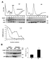
Ribosomal RACK1 recruits FMRP on translational machinery. RACK1 recruits FMRP on polyribosomes. (A), Amount of FMRP and rps6, examined by immunoblotting, on ribosomal fractions collected by polysome profiling conducted on SH-SY5Y cells overexpressing Myc-RACK1WT (WT) or Myc-RACK1DE (DE) proteins. (Lower) The graphic quantifies the amount of FMRP normalized to the level of rpS6 on ribosome and polyribosome fractions (from 2 to 8 fractions) measured by densitometry on bands related to immunoblottings of three independent experiments. All bar graphs represent the mean and S.D. (B) (Left) immunoblotting for FMRP, rpS6 and Myc antibodies on proteins eluted by histidine purification from SH-SY5Y cells overexpressing Myc-RACK1WT (WT) and Myc-RACK1DE (DE) proteins. The amount of FMRP purified from eluted imidazole RDE was more elevated than that purified with RWT, while the level of rpS6 was reduced in purified RDE. (Right), quantification of FMRP co-purified with WT or DE measured by densitometry on bands related to immunoblottings of three independent experiments. All bar graphs represent the mean and S.D. of FMRP level normalized to the amount of purified Myc-fusion proteins. Student’s T-test was used to calculate p values * < 0.01.
Figure 5
Figure 5
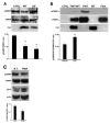
RACK1 and FMRP are part of the same complex. (A) RACK1 (red) and FMRP (green) in immunofluorescence experiments co-localize in the soma and partially along neurites of mouse embryonic hippocampal neurons. Inbox, the magnification of one neurite; the arrow indicates the granules where there is colocalization between RACK1 and FMRP. Scale bar: 10μm. Bar grapgh reproteing the Pearson’s coefficient of puncta along between RACK1 and FMRP measured by immunofluorescence experiments. The bar represents mean ± SD; n.70 puncta from three independent experiments. (B) Western blotting on eluted protein by RACK1-FMRP co-immunoprecipitation assay from mouse brain. Rabbit IgG was used as a negative control of co-immunoprecipitation. The images are representative of three independent experiments.
Figure 6
Figure 6
RACK1 up-regulation and PKC activation induce FMRP dephosphorylation at S499. (A) Western blotting for phosphorylated FMRP (pFMRP) and total FMRP (FMRP) in control and Myc-RACK1WT (WT) and Myc-RACK1DE (DE) cells. The bar graph summarizes the mean of the pFMRP level normalized to that of FMRP measured by densitometry on immunoblottings of three independent experiments. Student’s T-test was used to calculate p values * < 0.01. (B) The level of phosphorylated FMRP-GFP in control cells and in cells overexpressing FMRPWT-GFP, FMRPWT-GFP/RACK1WT-his-myc (FWT/WT), RACK1WT-his-myc (WT) and FMRPSA-GFP (FSA). As expected, the S499A mutation in FMRPWT abrogates its phosphorylation. The bar graph summarizes the mean GFP-FMRP level normalized to that of GFP-FMRP measured by densitometry on immunoblottings of three independent experiments. Student’s T-test was used to calculate p values * < 0.01. (C) Western blotting for phosphorylated FMRP (pFMRP) and total FMRP (FMRP) in PMA treated cells. The immunoblotting for ERK phosphorylation indicated the effect of PMA. The bar graph summarizes the mean of the pFMRP level normalized to that of tFMRP measured by densitometry on immunoblottings of three independent experiments. Student’s T-test was used to calculate p values * < 0.01.

References

    1. Simsek D., Tiu G.C., Flynn R.A., Byeon G.W., Leppek K., Xu A.F., Chang H.Y., Barna M. The Mammalian Ribo-interactome Reveals Ribosome Functional Diversity and Heterogeneity. Cell. 2017;169:1051–1065.e18. doi: 10.1016/j.cell.2017.05.022. - DOI - PMC - PubMed
    1. Imami K., Milek M., Bogdanow B., Yasuda T., Kastelic N., Zauber H., Ishihama Y., Landthaler M., Selbach M. Phosphorylation of the Ribosomal Protein RPL12/uL11 Affects Translation during Mitosis. Mol. Cell. 2018;72:84–98.e9. doi: 10.1016/j.molcel.2018.08.019. - DOI - PubMed
    1. Shi Z., Fujii K., Kovary K.M., Genuth N.R., Röst H.L., Teruel M.N., Barna M. Heterogeneous Ribosomes Preferentially Translate Distinct Subpools of mRNAs Genome-wide. Mol Cell. 2017;67:71–83.e7. doi: 10.1016/j.molcel.2017.05.021. - DOI - PMC - PubMed
    1. Ron D., Chen C.H., Caldwell J., Jamieson L., Orr E., Mochly-Rosen D. Cloning of an intracellular receptor for protein kinase C: A homolog of the beta subunit of G proteins. Proc. Natl. Acad. Sci. USA. 1994;91:839–843. doi: 10.1073/pnas.91.3.839. - DOI - PMC - PubMed
    1. Doan A.T., Huttenlocher A. RACK1 regulates Src activity and modulates paxillin dynamics during cell migration. Exp. Cell Res. 2007;313:2667–2679. doi: 10.1016/j.yexcr.2007.05.013. - DOI - PMC - PubMed

MeSH terms