. 2022 Oct 8;23(19):11931.
doi: 10.3390/ijms231911931.

Promyelocytic Leukemia Protein Potently Restricts Human Cytomegalovirus Infection in Endothelial Cells

Affiliations

Promyelocytic Leukemia Protein Potently Restricts Human Cytomegalovirus Infection in Endothelial Cells

Sven Seitz et al. Int J Mol Sci. .

Abstract

PML nuclear bodies (PML-NBs) are dynamic macromolecular complexes that mediate intrinsic immunity against viruses of different families, including human cytomegalovirus (HCMV). Upon HCMV infection, PML-NBs target viral genomes entering the nucleus and restrict viral immediate-early gene expression by epigenetic silencing. Studies from several groups performed in human fibroblast cells have shown that the major PML-NB components PML, Daxx, Sp100 and ATRX contribute to this repression in a cooperative manner. Their role for HCMV restriction in endothelial cells, however, has not yet been characterized although infected endothelium is thought to play a crucial role for HCMV dissemination and development of vascular disease in vivo. Here, we use conditionally immortalized umbilical vein endothelial cells (HEC-LTT) as a cell culture model to elucidate the impact of PML-NB proteins on lytic HCMV infection. Depletion of individual PML-NB proteins by lentiviral transduction showed a particularly strong antiviral effect of PML in HEC-LTT, compared to human fibroblasts. A closer characterization of this antiviral function revealed that PML may not only effectively inhibit HCMV immediate-early gene expression but also act at later steps of the viral replication cycle. At contrast, we surprisingly noted an antiviral behavior of Daxx in complementary approaches: Depletion of Daxx resulted in decreased viral gene expression, while overexpression of Daxx promoted HCMV infection. In summary, our data demonstrate a cell type-specific effect of PML-NB components on lytic HCMV infection and suggest an important role of PML in the inhibition of HCMV dissemination through infected endothelial cells.

Keywords: Daxx; HCMV; HEC-LTT; PML; PML nuclear bodies; cytomegalovirus; endothelial cells.


Conflict of interest statement

The authors declare no conflict of interest.

Figures

Figure 1
Figure 1
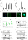
Characterization of PML-NBs in HEC-LTT. (A) Mean number of PML-NBs in the nuclei of HEC-LTT compared to HUVEC and HFF cells shown in a scatter plot. HEC-LTT cells were cultured in the presence of doxycycline (+Dox) or were deprived of doxycycline for one to seven days to induce growth arrest. HEC-LTT, HUVEC, and HFF cells were fixed and subjected to indirect immunofluorescence analysis of endogenous PML. Subsequent quantification of PML foci was performed in 50 cells per sample using maximum intensity projections of z-series images. (B) Localization of PML-NB proteins in HEC-LTT. HEC-LTT were cultured in presence (+Dox) or in absence (−Dox) of doxycycline for five days, before they were harvested and subjected to immunofluorescence analysis. Antibodies against PML and either Sp100, ATRX or Daxx were used. Representative images for the co-localization of PML-NB proteins are shown, which was analyzed in >50 cells per sample. (C) Western Blot analysis of PML-NB proteins in HEC-LTT and HFF cells. HEC-LTT were cultured in presence (+Dox) or absence (−Dox) of doxycycline for five days. HEC-LTT and HFF samples were harvested for Western blot analysis of PML, Sp100, Daxx, ATRX and β-actin as loading control. One representative Western blot experiment of ≥2 is shown.
Figure 2
Figure 2
Disruption of PML-NBs in HCMV-infected HEC-LTT. (A,B) Western Blot analysis of PML SUMOylation during HCMV infection. Growth-arrested HEC-LTT (A) and HFF (B) were infected with HCMV strain TB40/E at a MOI of 5. At indicated hours post infection (hpi), cells were harvested and subjected to Western blot analysis of endogenous PML as well as immediate–early (IE1), early (UL44), and late (MCP) phase viral proteins. β-actin was included as internal control. (C) Intracellular localization of PML and IE1 in infected HEC-LTT. Growth-arrested HEC-LTT were infected at a MOI of 5 with TB40/E. At different time points after infection, cells were fixed and stained by indirect immunofluorescence for PML and IE1. The depicted protein distributions were observed in >90% of analyzed cells (≥100 cells per sample).
Figure 3
Figure 3
Knockdown of individual PML-NB proteins in HEC-LTT and HFF cells. HEC-LTT (A,B) and HFF (C,D) were transduced with lentiviruses generated from an empty vector (shVector), a vector containing a non-functional shRNA (shControl) or vectors encoding shRNAs directed against PML, Sp100, Daxx and ATRX (shPML, shSp100, shATRX, shDaxx). Knockdown of the individual PML-NB proteins was confirmed by Western Blot (A,C) and immunofluorescence (B,D) analysis using indicated antibodies. The knockdown efficiency of the respective proteins was determined by analysis of >100 cells in immunofluorescence images and is indicated for HEC-LTT (B) and HFF (D) cells.
Figure 4
Figure 4
Analysis of HCMV IE gene expression in HEC-LTT and HFF with a knockdown of PML-NB proteins. (A,B) Effect of PML-NB protein knockdown on initiation of HCMV IE1 gene expression. HFF (A) and growth-arrested HEC-LTT (B) with a knockdown of PML-NB proteins (shPML, shSp100, shDaxx, shATRX) as well as control cells (shVector, shControl) were infected with 50 immediate–early units of TB40/E. At 24 hpi, the number of IE-expressing cells was determined by fluorescence microscopy using an IE1-specific antibody. Values are derived from triplicate samples and represent mean values ± SD. The results are representative for two independent experiments performed in triplicates. Datasets were analyzed by one-way-ANOVA to test for significant differences between the various cell populations. Post-hoc analysis was conducted to compare the individual datasets with shControl cells, which were used as control cells in all following experiments. Asterisks indicate statistically significant differences. ***, p < 0.001; **, p < 0.01; *, p < 0.05. (C,D) Effect of PML knockdown on nuclear translocation of HCMV particles. PML-knockdown and control HEC-LTT (growth-arrested) and HFF were infected with freshly harvested TB40/E. After 6 h, cells were fixed for immunofluorescence staining of the viral tegument protein pp150 and of viral IE1/IE2. An exemplary image of HEC-LTT shPML is shown (C). To determine the nuclear translocation efficiency of HCMV particles, pp150 signals at the nucleus were counted in 30 cells derived from three independent experiments and are shown as percentage of total intracellular pp150 signals. IE1/IE2 expression in the single cells is indicated by green coloring, grey color is used for cells in which no IE gene expression was initiated. Student’s t-test was performed for statistical analysis. ns, not significant (D). (E) Effect of Daxx depletion on HCMV infection in HEC-LTT. Growth-arrested control (shControl) and Daxx-knockdown (shDaxx) HEC-LTT were infected with TB40/E at a MOI of 3. At indicated times, the cells were subjected to Western Blot analysis of HCMV immediate–early (IE1) and early (UL44, UL84) proteins. β-actin was included as internal control. Levels of viral proteins were quantified via densitometric analyses using the Fusion Software (Vilber Lourmat GmbH). Protein levels were normalized to the internal control (β-actin) and are given as fold change. (F) Effect of Daxx depletion on HCMV infection in primary HUVEC. HUVEC were incubated with control lentiviruses (shControl) or lentiviruses encoding a shRNA against Daxx (shDaxx). Five days after transduction, the cells were infected with TB40/E at a MOI of 3. At indicated times after HCMV infection, cells were subjected to Western Blot analysis of Daxx as well as viral immediate–early (IE1) and early (UL44, UL84) proteins. β-actin was included as internal control. Levels of viral proteins were quantified as described in (E). (G) Effect of Daxx reintroduction on HCMV infection in HEC-LTT. Daxx-depleted HEC-LTT were transduced with control lentiviruses (shDaxx + control) or lentiviruses expressing Flag-tagged Daxx (shDaxx + Daxx) and selected with blasticidin, before they were mock infected or infected with TB40/E at a MOI of 3. At indicated times after infection, cells were subjected to Western Blot analysis of Daxx as well as viral immediate–early (IE1) and early (UL44, UL84) proteins. β-actin was included as internal control. Levels of viral proteins were quantified as described in (E).
Figure 5
Figure 5
MOI-independent inhibition of HCMV replication by PML in HEC-LTT. (A) Analysis of HCMV gene expression in PML-knockdown (shPML) HEC-LTT compared to control cells (shControl). Growth-arrested HEC-LTT were infected with TB40/E at a MOI of 1 and harvested at different times after infection. Western blot analysis was conducted to assess the abundances of viral immediate–early (IE), early (E), and late (L) proteins as indicated. β-actin was included as internal control. Levels of viral IE proteins at 24 hpi were quantified via densitometric analysis using the Fusion Software (Vilber Lourmat GmbH) and normalized to the internal control (β-actin). The mean densitometric values of three Western blots are given as fold-changes. (B) Quantification of HCMV DNA replication in PML-knockdown HEC-LTT compared to control cells. Growth-arrested PML-knockdown (shPML) and control (shControl) HEC-LTT were infected with TB40/E at a MOI of 0.05 or 0.5. At 5 dpi, intracellular DNA was extracted, and viral genome equivalents were determined via IE1-specific qPCR in relation to cellular albumin copy numbers. Values are derived from biological triplicates and represent mean values ± SD. Data sets were analyzed using unpaired Student’s t-tests. Asterisks indicate statistically significant differences. **, p < 0.01; ***, p < 0.001. (C) Quantification of virus progeny release from PML-knockdown HEC-LTT compared to control cells. Growth-arrested PML-knockdown and control HEC-LTT were infected with TB40/E at a MOI of 0.05 or 0.5. At 5 dpi, viral supernatants were harvested and subjected to protease K treatment followed by IE1-specific qPCR. Standard deviations of three biological replicates are shown. P-values were calculated using unpaired Student’s t-tests and are indicated by asterisks. *, p < 0.5.

References

    1. Griffiths P., Reeves M. Pathogenesis of human cytomegalovirus in the immunocompromised host. Nat. Rev. Microbiol. 2021;19:759–773. doi: 10.1038/s41579-021-00582-z. - DOI - PMC - PubMed
    1. Kagan K.O., Hamprecht K. Cytomegalovirus infection in pregnancy. Arch. Gynecol. Obstet. 2017;296:15–26. doi: 10.1007/s00404-017-4380-2. - DOI - PubMed
    1. Sinzger C., Digel M., Jahn G. Cytomegalovirus cell tropism. Curr. Top. Microbiol. Immunol. 2008;325:63–83. - PubMed
    1. Gerna G., Baldanti F., Revello M.G. Pathogenesis of human cytomegalovirus infection and cellular targets. Hum. Immunol. 2004;65:381–386. doi: 10.1016/j.humimm.2004.02.009. - DOI - PubMed
    1. Adler B., Sinzger C. Endothelial cells in human cytomegalovirus infection: One host cell out of many or a crucial target for virus spread? Thromb. Haemost. 2009;102:1057–1063. - PubMed

MeSH terms