Review
. 2022 Oct 8;23(19):11952.
doi: 10.3390/ijms231911952.

The Roles of Androgens in Humans: Biology, Metabolic Regulation and Health

Affiliations
Review

The Roles of Androgens in Humans: Biology, Metabolic Regulation and Health

Marià Alemany. Int J Mol Sci. .

Abstract

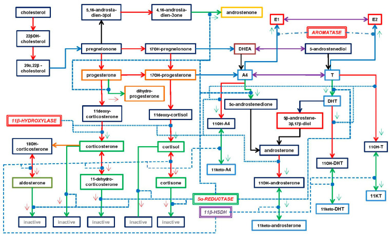
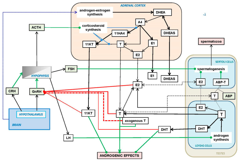
Androgens are an important and diverse group of steroid hormone molecular species. They play varied functional roles, such as the control of metabolic energy fate and partition, the maintenance of skeletal and body protein and integrity and the development of brain capabilities and behavioral setup (including those factors defining maleness). In addition, androgens are the precursors of estrogens, with which they share an extensive control of the reproductive mechanisms (in both sexes). In this review, the types of androgens, their functions and signaling are tabulated and described, including some less-known functions. The close interrelationship between corticosteroids and androgens is also analyzed, centered in the adrenal cortex, together with the main feedback control systems of the hypothalamic-hypophysis-gonads axis, and its modulation by the metabolic environment, sex, age and health. Testosterone (T) is singled out because of its high synthesis rate and turnover, but also because age-related hypogonadism is a key signal for the biologically planned early obsolescence of men, and the delayed onset of a faster rate of functional losses in women after menopause. The close collaboration of T with estradiol (E2) active in the maintenance of body metabolic systems is also presented Their parallel insufficiency has been directly related to the ravages of senescence and the metabolic syndrome constellation of disorders. The clinical use of T to correct hypoandrogenism helps maintain the functionality of core metabolism, limiting excess fat deposition, sarcopenia and cognoscitive frailty (part of these effects are due to the E2 generated from T). The effectiveness of using lipophilic T esters for T replacement treatments is analyzed in depth, and the main problems derived from their application are discussed.

Keywords: anabolic steroids; androgens; dehydroepiandrosterone; dihydrotestosterone; estradiol; metabolic regulation; metabolic syndrome; senescence; testosterone; testosterone replacement therapy.


Conflict of interest statement

The author declares no conflict of interest.

Figures

Figure 1
Figure 1
Main androgen sytntheis pathways. This figure represents the physiological molecular species secreted in/by adrenal glands (intermediate and cortical layers), testicles, ovaries and the brain, as well by a number of other organs or tissues with a critical participation in these processes (e.g., skin, liver, adipose tissue). Since the synthesis of androgens (especially in the adrenal glands) is closely related to the two parallel corticosteroid synthesis pathways (they share location and a few enzyme activities), the start of these paths has been indicated in green squares. This includes the estrogens, which metabolism is much more closely intertwined with that of the main androgens. The 16-[estriol] and 11-hydroxylative pathways, as well as the catechol-estrogen specific pathway, have been included only as annotations in green labels. Black arrows show the enzyme-driven changes between molecular species; two-headed arrows show reactions that are potentially bidirectional. The main androgen molecule borders are red, violet in those sharing androgen and estrogen capabilities and blue in the fully estrogenic molecules; the progestogen borders are marked in yellow, and the androgenic pheromone species are in grey. The remaining molecules (black borders) may show a limited (if any) androgen receptor binding ability. The enzymes intervening in the reactions depicted are listed below the figure. They are presented in borderless pale blue rectangles in contact with the corresponding black arrows; the letters are in brown for mitochondrial and black for microsomal (and other location) enzymes.
Figure 1
Figure 1
Main androgen sytntheis pathways. This figure represents the physiological molecular species secreted in/by adrenal glands (intermediate and cortical layers), testicles, ovaries and the brain, as well by a number of other organs or tissues with a critical participation in these processes (e.g., skin, liver, adipose tissue). Since the synthesis of androgens (especially in the adrenal glands) is closely related to the two parallel corticosteroid synthesis pathways (they share location and a few enzyme activities), the start of these paths has been indicated in green squares. This includes the estrogens, which metabolism is much more closely intertwined with that of the main androgens. The 16-[estriol] and 11-hydroxylative pathways, as well as the catechol-estrogen specific pathway, have been included only as annotations in green labels. Black arrows show the enzyme-driven changes between molecular species; two-headed arrows show reactions that are potentially bidirectional. The main androgen molecule borders are red, violet in those sharing androgen and estrogen capabilities and blue in the fully estrogenic molecules; the progestogen borders are marked in yellow, and the androgenic pheromone species are in grey. The remaining molecules (black borders) may show a limited (if any) androgen receptor binding ability. The enzymes intervening in the reactions depicted are listed below the figure. They are presented in borderless pale blue rectangles in contact with the corresponding black arrows; the letters are in brown for mitochondrial and black for microsomal (and other location) enzymes.
Figure 2
Figure 2
Types of human androgens. The androgen molecular species (or groups of them) show similar chemical structures, are synthesized in a number of different tissues (but mainly in adrenal glands and the gonads) and elicit physiological effects that are largely complementary. DHEA: dehydroepiandrosterone; T: testosterone; AcT: 17β-acyl-T, a group of esters; KTs: 11-keto-androgens (essentially derived from T, DHT and A4; the 11-keto forms are more active than the 11-hydroxyl ones); DHT: dihydrotestosterone; A4: 4-androstenedione; AP: androgenic pheromones; EA: estrogenic androgens (i.e., they bind to the ER and AR); Ane: androsterone—a catabolite of T—which is an agonist of the farnesoid receptor, acting in the regulation of bile acid signaling. There are many other androgen catabolism products and intermediate molecular species of the androgen metabolism, which specialized functions have not been described in depth, but have been studied as pharmacological subjects, metabolic markers or substrates for synthetic hormone production. The androgens susceptible of aromatization are marked with green benzene icons. Small red arrows point to the distinguishing structural features of the different groups of androgens in comparison with T taken as the standard and best-known androgen.
Figure 3
Figure 3
Functional structure of the androgen receptor (AR). The diagram shows the two distinct parts of the protein chain, joined at a flexible joint (hinge). The longest arm contains the N-terminal domain, incorporating the AF1 (activation function 1) binding sites, as well as the DNA-binding domain and a short sequence (nuclear localization signal) needed for the nuclear transport of the AR. The shorter arm (C-terminal domain) contains the AF2 (activation function 2) binding site. This domain contains a key binding niche, the LBP (ligand-binding pocket), in the core of AF2.
Figure 4
Figure 4
Main shared enzyme activities in the control of glucocorticoid and androgen metabolism in the adrenal gland. Critical role of four key enzymes: aromatase, 5α-reductase, 11β-hydroxylase and 11β-hydroxysteroid dehydrogenase, in the activity and regulation of the main synthesis interrelationships between glucocorticoids, androgens and estrogens. Main glucocorticoids: green-rimmed rectangles (aldosterone: olive green); androgens: dark blue (DHEA: brown) and estrogens: red. The reductive reactions are green arrows, oxidative reactions; red arrows; coenzyme-dependent reversible oxidation-reduction reactions; violet double-pointed arrows (i.e., oxidative or reductive, depending on the tissue redox status). The enzymes are oxidative in red capital letters, reductive in green and equilibrium-oxidation-reductive in violet. Only the four selected key enzymes are listed; those intervening in androgen metabolism have been already described in detail in Figure 1. The actions of enzymes are separately described. The reactions catalyzed have been marked with the color corresponding to the effect elicited: red—oxidation, green—reduction; additional small arrows mark the effects on the product of the overall reaction. Enzyme actions are marked by blue discontinuous lines: aromatase is the point-dash line; 5α-reductase is the dashed line; 11β-hydroxylase is the points line; and 11β-HSDH is the thinner dashed line. Mixed reactions (such as that of aromatase) or actions carried out by non-oxidative-reductive enzymes have been left in black. As explained in the text, the formation of KT and similar compounds are oxidative-activating processes for androgens, but oxidative-inhibiting processes for glucocorticoids. In a reverse way, the action of 5α-reductase enhances the synthesis of DHT and androstenone, but inactivates the oxidized forms of glucocorticoids. Finally, aromatase irreversibly converts A4 or T into estrogens, thus, leaving a narrow (and critically controlled) path for the production of estrogens. In fact, a few key enzymes and a varying metabolic oxidative or reductive ambiance may deeply affect the outcome of the main classes of steroid hormones in a coordinate and partly auto-regulating mechanism.
Figure 5
Figure 5
Regulation of the androgen (and estrogen) synthesis by the adrenal cortex and testes. Abbreviations: GnRH: gonadotropin-releasing hormone; CRH: corticotropin-releasing hormone; ACTH: corticotropin; (gonadotropins) FSH: follicle-stimulating hormone; LH: luteotropic hormone; E1: estrone; E2: 3,17β-estradiol; ABP: androgen-binding protein. The other abbreviations have been described in Figure 2. The solid black arrows show paths or relationships. The dotted black line depicts the brain modulation of steroid hormone synthesis by the brain through other additional means (i.e., nervous signals and non-steroidal hormones). The dashed black lines indicate the possibility of direct interchange of pools between tissue and blood. The green lines show stimulatory/activating effects, and the red lines inhibitory/deactivating effects. The effects of exogenous T (i.e., administered as a drug) on GnRH functions overall have been presented as a red dashed line to somehow differentiate it from the tissue-released T.

References

    1. Comitato R., Saba A., Turrini A., Arganini C., Virgili F. Sex hormones and macronutrient metabolism. Crit. Rev. Food Sci. Nutr. 2015;55:227–241. doi: 10.1080/10408398.2011.651177. - DOI - PMC - PubMed
    1. Giudicelli Y., Dieudonne M.N., Lacasa D., Pasquier Y.N., Pecquery R. Modulation by sex hormones of the membranous transducing system regulating fatty acid mobilization in adipose tissue. Prostaglandins Leukot. Essent. Fat. Acids. 1993;48:91–100. doi: 10.1016/0952-3278(93)90015-O. - DOI - PubMed
    1. Remesar X., Alemany M. Dietary energy partition: The central role of glucose. Int. J. Mol. Sci. 2020;21:7729. doi: 10.3390/ijms21207729. - DOI - PMC - PubMed
    1. Stumpf W.E. Steroid hormones and the cardiovascular system: Direct actions of estradiol, progesterone, testosterone, gluco- and mineralcorticoids, and soltriol (vitamin D) on central nervous regulatory and peripheral tissues. Experientia. 1990;46:13–25. doi: 10.1007/BF01955408. - DOI - PubMed
    1. Fietta P., Fietta P., Delsante G. Central nervous system effects of natural and synthetic glucocorticoids. Psychiatry Clin. Neurosci. 2009;63:613–622. doi: 10.1111/j.1440-1819.2009.02005.x. - DOI - PubMed

LinkOut - more resources