. 2022 Oct 9;23(19):12004.
doi: 10.3390/ijms231912004.

Saposin C, Key Regulator in the Alpha-Synuclein Degradation Mediated by Lysosome

Affiliations

Saposin C, Key Regulator in the Alpha-Synuclein Degradation Mediated by Lysosome

Clara Ruz et al. Int J Mol Sci. .

Abstract

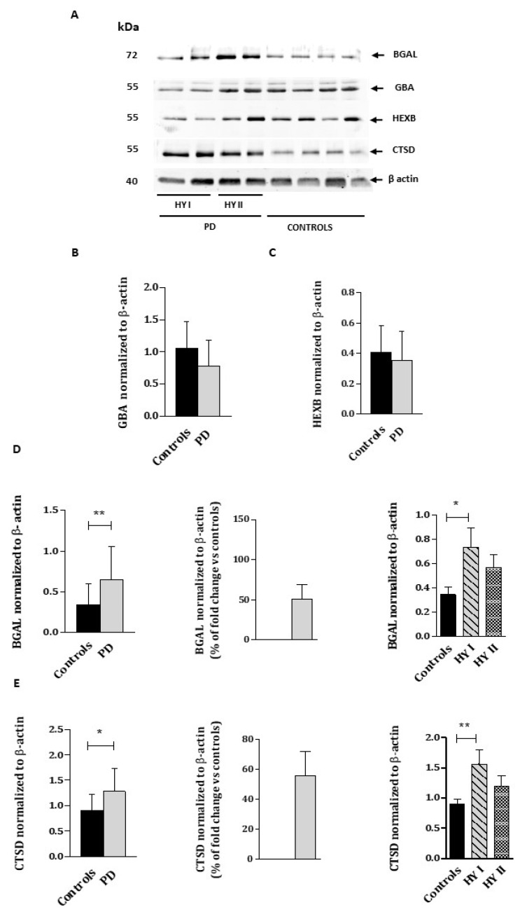
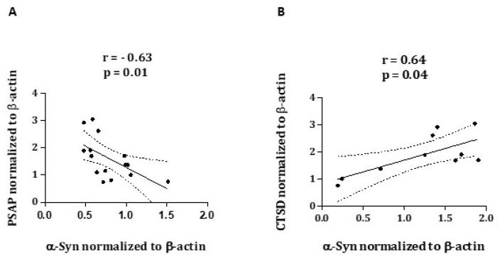
Lysosomal dysfunction has been proposed as one of the most important pathogenic molecular mechanisms in Parkinson disease (PD). The most significant evidence lies in the GBA gene, which encodes for the lysosomal enzyme β-glucocerebrosidase (β-GCase), considered the main genetic risk factor for sporadic PD. The loss of β-GCase activity results in the formation of α-synuclein deposits. The present study was aimed to determine the activity of the main lysosomal enzymes and the cofactors Prosaposin (PSAP) and Saposin C in PD and healthy controls, and their contribution to α-synuclein (α-Syn) aggregation. 42 PD patients and 37 age-matched healthy controls were included in the study. We first analyzed the β-GCase, β-galactosidase (β-gal), β-hexosaminidase (Hex B) and Cathepsin D (CatD) activities in white blood cells. We also measured the GBA, β-GAL, β-HEX, CTSD, PSAP, Saposin C and α-Syn protein levels by Western-blot. We found a 20% reduced β-GCase and β-gal activities in PD patients compared to controls. PSAP and Saposin C protein levels were significantly lower in PD patients and correlated with increased levels of α-synuclein. CatD, in contrast, showed significantly increased activity and protein levels in PD patients compared to controls. Increased CTSD protein levels in PD patients correlated, intriguingly, with a higher concentration of α-Syn. Our findings suggest that lysosomal dysfunction in sporadic PD is due, at least in part, to an alteration in Saposin C derived from reduced PSAP levels. That would lead to a significant decrease in the β-GCase activity, resulting in the accumulation of α-syn. The accumulation of monohexosylceramides might act in favor of CTSD activation and, therefore, increase its enzymatic activity. The evaluation of lysosomal activity in the peripheral blood of patients is expected to be a promising approach to investigate pathological mechanisms and novel therapies aimed to restore the lysosomal function in sporadic PD.

Keywords: Cathepsin D; PSAP; Parkinson’s disease; lysosomal dysfunction; β-glucocerebrosidase.


Conflict of interest statement

The authors declare no conflict of interest.

Figures

Figure 1
Figure 1
Lysosomal enzymatic activity of β-GCase (A), Hex B (B), β-gal (C), and CatD (D) in WBC from PD patients compared to healthy controls. The percentage of fold change over controls was calculated as follows: (PD value—control activity average)/control activity average × 100. Data show mean ± SEM. * p < 0.05; *** p < 0.001.
Figure 2
Figure 2
Immunoblot analysis of GBA, BGAL, HEXB, and CTSD proteins (A) from the WBC lysates of PD patients and healthy controls. Molecular weight markers are indicated in kilodaltons. Quantification of GBA (B), BGAL (C), HEXB (D), and CTSD (E) protein levels normalized to loading control β-actin. HYI and HYII corresponded PD patients at Hoehn and Yahr stages I and II. Quantifications were based on four independent experiments (N = 16 per group). The percentage of fold change over controls was calculated as follows: (PD value– control protein average)/control protein average × 100. Values represent mean ± SEM. * p < 0.05, ** p < 0.01.
Figure 3
Figure 3
Immunoblot analysis of PSAP, Saposin C (A), and α-Syn (B) protein levels from the WBC lysates of PD patients and healthy controls. Molecular weight markers are indicated in kilodaltons. The quantification of PSAP (C), Saposin C (D), and α-Syn (E) protein levels normalized to loading control β-actin. HYI and HYII correspond to PD patients at Hoehn and Yahr stages I and II. Quantifications were based on four independent experiments (n = 16 per group). The percentage of fold change over controls was calculated as follows: (PD value—control protein average)/control protein average × 100. Values represent mean ± SEM. * p < 0.05, ** p < 0.01.
Figure 4
Figure 4
Correlation analysis of PSAP and α-Syn (A) and CTSD and α-Syn (B), obtained from Western blots from 15 and 10 PD patients, respectively.

References

    1. Tysnes O.B., Storstein A. Epidemiology of Parkinson’s disease. J. Neural. Transm. 2017;124:901–905. doi: 10.1007/s00702-017-1686-y. - DOI - PubMed
    1. Klingelhoefer L., Reichmann H. Parkinson’s disease as a multisystem disorder. J. Neural Transm. 2017;124:709–713. doi: 10.1007/s00702-017-1692-0. - DOI - PubMed
    1. Bandres-Ciga S., Diez-Fairen M., Kim J.J., Singleton A.B. Genetics of Parkinson’s disease: An introspection of its journey towards precision medicine. Neurobiol. Dis. 2020;137:104782. doi: 10.1016/j.nbd.2020.104782. - DOI - PMC - PubMed
    1. Ciechanover A., Kwon Y.T. Degradation of misfolded proteins in neurodegenerative diseases: Therapeutic targets and strategies. Exp. Mol. Med. 2015;47:e147. doi: 10.1038/emm.2014.117. - DOI - PMC - PubMed
    1. Cook C., Stetler C., Petrucelli L. Disruption of protein quality control in Parkinson’s disease. Cold Spring. Harb. Perspect Med. 2012;2:a009423. doi: 10.1101/cshperspect.a009423. - DOI - PMC - PubMed