Review
. 2022 Sep 22;11(19):5565.
doi: 10.3390/jcm11195565.

The Pancreas and Known Factors of Acute Pancreatitis

Affiliations
Review

The Pancreas and Known Factors of Acute Pancreatitis

Julia Walkowska et al. J Clin Med. .

Abstract

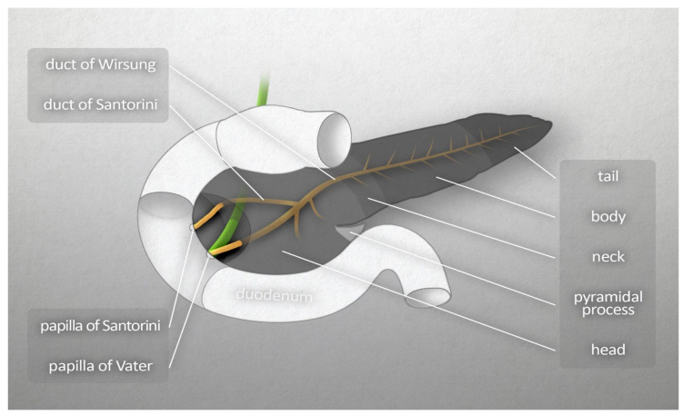
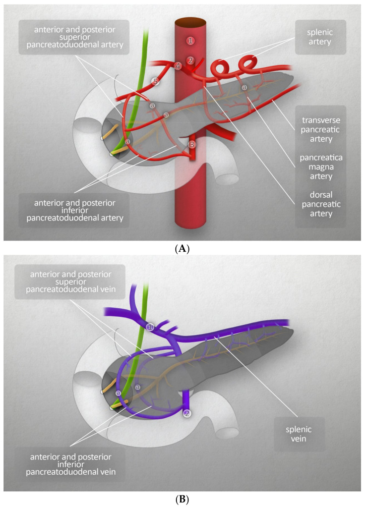
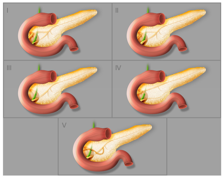
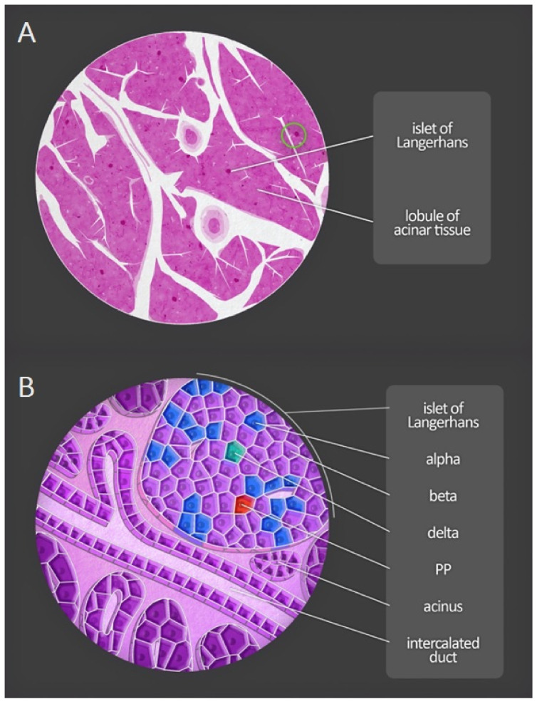
Pancreatitis is regarded by clinicians as one of the most complicated and clinically challenging of all disorders affecting the abdomen. It is classified on the basis of clinical, morphological, and histological criteria. Causes of acute pancreatitis can easily be identified in 75-85% of patients. The main causes of acute, recurrent acute, and chronic pancreatitis are gallstone migration and alcohol abuse. Other causes are uncommon, controversial, or unexplained. For instance, cofactors of all forms of pancreatitis are pancreas divisum and hypertriglyceridemia. Another factor that should be considered is a complication of endoscopic retrograde cholangiopancreatography: post-endoscopic retrograde cholangiopancreatography acute pancreatitis. The aim of this study is to present the known risk factors for acute pancreatitis, beginning with an account of the morphology, physiology, and development of the pancreas.

Keywords: pancreas; pancreatitis; risk factors; surgery.


Conflict of interest statement

The authors declare no conflict of interest.

Figures

Figure 1
Figure 1
Macroscopic anatomy of the human pancreas.
Figure 2
Figure 2
(A) Major blood vessels of the pancreas- arteries: (1) abdominal aorta, (2) celiac trunk, (3) superior mesenteric artery, (4) common hepatic artery, (5) gastroduodenal artery, (a) anastomotic branch. (B) Major blood vessels of the pancreas-veins: (1) hepatic portal vein, (2) superior mesenteric vein, (a) anastomotic branch.
Figure 3
Figure 3
Different types of pancreatic duct configuration: (I) bifid configuration with pancreatic duct as the dominant duct, (II) bifid configuration with accessory pancreatic duct as the dominant duct, (III) rudimentary nondraining or absent accessory pancreatic duct, (IV) pancreas divisum, (V) ansa pancreatica.
Figure 4
Figure 4
Microscopic anatomy of the human pancreas. (A) Magnification of a portion of the pancreas shows both lobules and pancreatic islets. (B) At greater magnification, acini and excretory ducts are visible. Different types of endocrine cells composing the pancreatic islets can be distinguished by immunofluorescent staining. PP—pancreatic polypeptide cells.

Similar articles

Cited by

References

    1. Dimitriou I., Katsourakis A., Nikolaidou E., Noussios G. The Main Anatomical Variations of the Pancreatic Duct System: Review of the Literature and Its Importance in Surgical Practice. J. Clin. Med. Res. 2018;10:370–375. doi: 10.14740/jocmr3344w. - DOI - PMC - PubMed
    1. Suda K., Nobukawa B., Takase M., Hayashi T. Pancreatic segmentation on an embryological and anatomical basis. J. Hepato-Biliary-Pancreat. Surg. 2006;13:146–148. doi: 10.1007/s00534-005-1039-3. - DOI - PubMed
    1. Dolenšek J., Rupnik M.S., Stožer A. Structural similarities and differences between the human and the mouse pancreas. Islets. 2015;7:e1024405. doi: 10.1080/19382014.2015.1024405. - DOI - PMC - PubMed
    1. Röder P.V., Wu B., Liu Y., Han W. Pancreatic regulation of glucose homeostasis. Exp. Mol. Med. 2016;48:e219. doi: 10.1038/emm.2016.6. - DOI - PMC - PubMed
    1. Arsenijevic T., Perret J., van Laethem J.L., Delporte C. Aquaporins involvement in pancreas physiology and in pancreatic diseases. Int. J. Mol. Sci. 2019;20:5052. doi: 10.3390/ijms20205052. - DOI - PMC - PubMed

LinkOut - more resources