Case Reports
. 2022 Sep 26;11(19):5684.
doi: 10.3390/jcm11195684.

Subcapsular Liver Hematoma-A Life-Threatening Condition in Preterm Neonates-A Case Series and Systematic Review of the Literature

Affiliations
Case Reports

Subcapsular Liver Hematoma-A Life-Threatening Condition in Preterm Neonates-A Case Series and Systematic Review of the Literature

Paraskevi Liakou et al. J Clin Med. .

Abstract

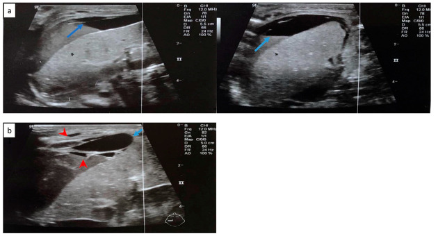
The subcapsular hematoma (SLH) of the liver is a rare finding in living infants. The clinical presentation of rupture is non-specific, with the signs of hypovolemic shock dominating. The causes are several, with prematurity, trauma and sepsis playing a leading role in the creation of an SHL. Umbilical vein catheterization and an increased bleeding tendency have also been associated with this usually fatal diagnosis. Abdominal ultrasonography, among other imaging methods, comprises the gold standard examination for early diagnosis. It should be differentiated from other possible causes of shock, such as sepsis and intraventricular hemorrhage, which have similar clinical presentation. We report a case series of three very low birth weight preterms (VLBW), with an SHL, during the first days of life, one of which survived from this usually catastrophic condition. A comprehensive review of the literature regarding this clinical entity was also conducted. A high index of suspicion is essential for early identification of such a case, with conservative or surgical treatment being the way to go.

Keywords: neonates; preterm neonates; subcapsular liver hematoma; very-low-birth-weight.


Conflict of interest statement

The authors declare no conflict of interest.

Figures

Figure 1
Figure 1
Hemostatic profile of the patient at the day of clinical deterioration, as depicted by ROTEM EXTEM and FIBTEM graphic representation and numerical results. Abbreviations: CT, clotting time (min, green colour); CFT, clot formation time (min, pink colour); A5 and A10, clot amplitude at 5 and 10 min (mm); MCF: maximum clot firmness (mm, blue colour); LI60, lysis index at 60 min (%).
Figure 2
Figure 2
Abdominal ultrasonography depicting a cystic formation 2.1 cm × 0.5 cm in the anterior portion of the right liver, with anechoic characteristics (blue arrow).
Figure 3
Figure 3
Radiography of the neonate on the day of deterioration. Non-specific signal characteristics in the abdomen, such as distention and poor distribution of bowel gas. A clavicular fracture clavicle is noted on the right (red arrowhead).
Figure 4
Figure 4
Hemostatic profile of the patient on the day of clinical deterioration, as illustrated by ROTEM EXTEM and FIBTEM graphic representation and numerical results. Abbreviations: CT, clotting time (min, green colour); CFT, clot formation time (min, pink colour); A5 and A10, clot amplitude at 5 and 10 min (mm); MCF: maximum clot firmness (mm); LI60, lysis index at 60 min (%).
Figure 5
Figure 5
(a) Free perihepatic fluid collection (blue arrow) in abdominal ultrasound. (b) Abdominal ultrasound depicting subcapsular hematoma of the liver (blue arrow) and hemorrhagic collections in the peritoneal cavity (red arrowhead). Normal liver parenchyma is noted with asterisk (*).
Figure 6
Figure 6
Radiography of the infant on the day of deterioration, with non-specific signal characteristics in abdomen, such as distended abdomen and poor distribution of bowel gas.
Figure 7
Figure 7
Hemostatic profile of patient on the day of clinical deterioration, as depicted by ROTEM EXTEM and FIBTEM graphic representation and numerical results. Abbreviations: CT, clotting time (min, green colour); CFT, clot formation time (min, pink colour); A5 and A10, clot amplitude at 5 and 10 min (mm); MCF: maximum clot firmness (mm, blue colour); LI60, lysis index at 60 min (%).
Figure 8
Figure 8
(a) Abdominal ultrasonography depicting hypoechogenic intrahepatic irregular lesion on the right lobe (3.4 cm × 1 cm) (blue arrow); and (b) abdominal ultrasonography depicting the calcific deposits (1 cm × 1 cm) (blue arrow) at the right lobe of the liver. Normal liver parenchyma is noted with asterisk (*).
Figure 9
Figure 9
(a) Chest and abdominal radiography depicting the abnormal position of umbilical venous catheter in the liver (red arrowhead); and (b) chest and abdominal radiography depicting air in the branches of right portal vein and hepatic parenchyma (red arrowhead).

References

    1. Purisch S.E., Gyamfi-Bannerman C. Epidemiology of preterm birth. Semin. Perinatol. 2017;41:387–391. doi: 10.1053/j.semperi.2017.07.009. - DOI - PubMed
    1. Theyskens C., Dams A. Successful Treatment of Liver Rupture in a Premature Infant. Adv. Neonatal Care Off. J. Natl. Assoc. Neonatal Nurses. 2011;11:25–26. doi: 10.1097/ANC.0b013e318206d8a3. - DOI - PubMed
    1. Anjay M.A., Sasidharan C.K., Anoop P. Hepatic subcapsular hematoma: Two neonates with disparate presentations. Pediatrics Neonatol. 2012;53:144–146. doi: 10.1016/j.pedneo.2012.01.013. - DOI - PubMed
    1. Park J.H., Cho W., Kwon M.A. Unexpected extensive hemorrhage from a subcapsular hematoma of the liver during emergent laparotomy in a premature neonate. Saudi Med. J. 2019;40:836–839. doi: 10.15537/smj.2019.8.24376. - DOI - PMC - PubMed
    1. Singer D.B., Neave C., Oyer C.E., Pinar H. Hepatic Subcapsular Hematomas in Fetuses and Neonatal Infants. Pediatric Dev. Pathol. 1999;2:215–220. doi: 10.1007/s100249900116. - DOI - PubMed

Publication types

LinkOut - more resources