Review
. 2022 Oct 7;11(19):5921.
doi: 10.3390/jcm11195921.

A Comprehensive Review of Pediatric Acute Encephalopathy

Affiliations
Review

A Comprehensive Review of Pediatric Acute Encephalopathy

George Imataka et al. J Clin Med. .

Abstract

Acute encephalopathy typically affects previously healthy children and often results in death or severe neurological sequelae. Acute encephalopathy is a group of multiple syndromes characterized by various clinical symptoms, such as loss of consciousness, motor and sensory impairments, and status convulsions. However, there is not only localized encephalopathy but also progression from localized to secondary extensive encephalopathy and to encephalopathy, resulting in a heterogeneous clinical picture. Acute encephalopathy diagnosis has advanced over the years as a result of various causes such as infections, epilepsy, cerebrovascular disorders, electrolyte abnormalities, and medication use, and new types of acute encephalopathies have been identified. In recent years, various tools, including neuroradiological diagnosis, have been developed as methods for analyzing heterogeneous acute encephalopathy. Encephalopathy caused by genetic abnormalities such as CPT2 and SCN1A is also being studied. Researchers were able not only to classify acute encephalopathy from image diagnosis to typology by adjusting the diffusion-weighted imaging/ADC value in magnetic resonance imaging diffusion-weighted images but also fully comprehend the pathogenesis of vascular and cellular edema. Acute encephalopathy is known as a very devastating disease both medically and socially because there are many cases where lifesaving is sometimes difficult. The overall picture of childhood acute encephalopathy is becoming clearer with the emergence of the new acute encephalopathies. Treatment methods such as steroid pulse therapy, immunotherapy, brain hypothermia, and temperature control therapy have also advanced. Acute encephalopathy in children is the result of our predecessor's zealous pursuit of knowledge. It is reasonable to say that it is a field that has advanced dramatically over the years. We would like to provide a comprehensive review of a pediatric acute encephalopathy, highlighting advancements in diagnosis and treatment based on changing disease classification scenarios from the most recent clinical data.

Keywords: acute encephalopathy; brain hypothermia; convulsions; pediatrics.


Conflict of interest statement

The authors declare no conflict of interest.

Figures
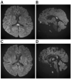
Figure 1
Figure 1
Imaging Characteristics of MERS. (A,B): A Horizontal/B sagittal section was performed on an 8-year-old boy who had an MRI on the third day of fever due to impaired consciousness and unable to recognize his own name. DWI showed an abnormal high signal in the cerebral corpus callosum: WBC 24800, CRP 7.84, Na 133, CL 95, ferritin 119.9, IL-6 171 EEG showed high amplitude slow waves in the occipital region. After 3 days of steroid pulse therapy, the fever resolved and consciousness improved. No sequelae. No causative organism or virus could be identified. (C,D): On the third day of vomiting and fever, he was hospitalized because he could no longer talk to his mother and could not look at her. He had diarrhea and was positive for rotavirus antigen in stool. In the bilateral frontal and occipital regions, EEG revealed persistent high amplitude slow waves. He was diagnosed with MERS on the fifth day after a diffusion-weighted MRI revealed an abnormally high signal in the corpus callosum. mPSL steroid pulse therapy was administered for three days, his level of consciousness improved, and both EEG and MRI were normalized.
Figure 2
Figure 2
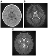
Imaging Characteristics of ANE. (A): An 11-month-old boy was admitted to the hospital after experiencing fever and vomiting. He was given a cold medicine prescription and sent home, but the next day, after a 3 min febrile convulsion, his loss of consciousness lasted 12 h, and a second 3 min convulsion was noted, so a CT was performed. He was diagnosed with ANE after a CT scan of the brain revealed abnormalities in the bilateral thalamus. (B): A 4-year-old boy visited the hospital with a high fever, vomiting, impaired consciousness, and convulsions. The rapid influenza A antigen test was positive, and MRI indicated abnormal signals in the bilateral thalamus not only on diffusion-weighted but also on T2 images, leading to the diagnosis of ANE. The patient was admitted to the intensive care unit immediately after being diagnosed with ANE. He was given cerebral sedation with high-dose barbital therapy and cerebral hypothermia at 34.5 °C for 48 h, which was followed by TTM as temperature control therapy, IVIG high-dose therapy, and mPSL steroid pulse therapy. Mitochondrial cocktail therapy was used in combination with 2 months after onset; the patient was able to walk, and 4 months later, his speech function had recovered to the same level as before the onset. (C): A 1-year and 2-month-old girl was admitted to the hospital with fever and partial seizures. After an MRI the next day, the T2-weighted image showed abnormal signals in the bilateral thalamus and diagnosed ANE. She was treated in the intensive care unit with 72 h 34.5 °C brain hypothermia, steroid pulse therapy, IVIG, and mitochondrial cocktail therapy. The patient was given cerebral sedation with high-dose barbital therapy and she was treated with dextromethorphan, which saved her life, but she was left with severe neurological sequelae.
Figure 3
Figure 3
Imaging characteristics of AESD. (A) An 11-month-old boy who was admitted to the hospital with a high fever and 15 min seizure congestion of the right upper and lower extremities. The seizures stopped after the administration of midazolam. Thereafter, there was transient Todd’s palsy of the right upper and lower extremities. Brain MRI was normal. The fever resolved 3 days later and a rash appeared, which was clinically diagnosed as HHV-6 infection. The second diffusion-weighted brain MRI showed a bright tree appearance sign predominantly on the left side, diagnosing AESD. mPSL 30 mg/kg 3 days pulse therapy was administered. At the age of 6, he entered a regular elementary school, but his language skills were mildly poor. (B) A 3-year and 3-month-old girl. She has a 1 h febrile convulsion superimposed on fever. Midazolam brought the convulsions to a halt. The next day, she remained listless and was monitored with intravenous fluids; on the eighth day, she experienced a cluster of short convulsions in her limbs. Diffusion-weighted brain MRI revealed bilateral subcortical white matter predominance with bright tree appearance and an AESD diagnosis. Then, 48 h of mild cerebral hypothermia at 35.5 °C, steroid pulse therapy, and mitochondrial rescue therapy were performed. Six years after onset, she is living a normal fourth-grade elementary school life with no sequelae in terms of motor, language, or academic performance. (C) A 1-year and 7-month-old boy. After 4 days of febrile convulsive seizures, the fever subsided and a rash appeared; he was clinically diagnosed with HHV-6 infection. Multiple convulsive seizures lasting a few minutes were observed 5 days later. Slow waves were detected in the frontal and occipital regions of the EEG. Diffusion-weighted brain MRI showed an abnormally high signal in subcortical white matter and diagnosed AESD. mPSL pulse therapy and vitamin cocktail therapy were started. Body temperature was maintained at 35.5–36.0 TTM for 5 days The disease has been present for over two and a half years, and the child is now over 4 years old. There are no neurological sequelae and both language and motor functions are age-appropriate. (D) A 1-year-old boy with a fever of 39 °C and spontaneous convulsions that stopped spontaneously before reaching the hospital; 4 days later, he presents with two 3-min generalized convulsions and is rushed to the emergency room with no recovery of consciousness. He was admitted directly to the ICU, sedated with Rabonar, and given 48 h of mild cerebral hypothermia at 35 °C. Steroid pulse therapy was also administered. Thereafter, the temperature was kept at 36 °C, and the patient was transferred from the ICU to the general ward on the eighth day. On the same day, a brain MRI showed an abnormally high signal on diffusion-weighted images with bilateral frontal lobe predominance, and a diagnosis of AIEF-type AESD was made. Rehabilitation was continued until he was over 2 years old. After 1 year of onset, both his motor and language functions have recovered to the level of his age.
Figure 4
Figure 4
Imaging Characteristics of PRES. (A,B): A 13-year-old boy underwent skin graft surgery due to severe burns all over his body. He was on ventilatory management and sedatives for a long period of time postoperatively. As his generalized sepsis improved, his anesthetic was reduced and he was awakened; after 50 days, his consciousness improved completely; on day 51, he complained that “everything I see is white and I can’t see anything.” He then had a severe headache. His blood pressure was 150/89 mmHg and he had hypertension. Brain MRI scan showed an abnormal high signal in bilateral occipital areas on T2-weighted (A) and FLAIR (B) images, and he was diagnosed with PRES.

References

    1. Slooter A.J.C., Otte W.M., Devlin J.W., Arora R.C., Bleck T.P., Claassen J., Duprey M.S., Ely E.W., Kaplan P.W., Latronico N., et al. Updated nomenclature of delirium and acute encephalopathy: Statement of ten Societies. Intensive Care Med. 2020;46:1020–1022. doi: 10.1007/s00134-019-05907-4. - DOI - PMC - PubMed
    1. Mizuguchi M., Ichiyama T., Imataka G., Okumura A., Goto T., Sakuma H., Takanashi J.I., Murayama K., Yamagata T., Yamanouchi H., et al. Guidelines for the diagnosis and treatment of acute encephalopathy in childhood. Brain Dev. 2021;43:2–31. doi: 10.1016/j.braindev.2020.08.001. - DOI - PubMed
    1. Hoshino A., Saitoh M., Oka A., Okumura A., Kubota M., Saito Y., Takanashi J., Hirose S., Yamagata T., Yamanouchi H., et al. Epidemiology of acute encephalopathy in Japan, with emphasis on the association of viruses and syndromes. Brain Dev. 2012;34:337–343. doi: 10.1016/j.braindev.2011.07.012. - DOI - PubMed
    1. Kasai M., Shibata A., Hoshino A., Maegaki Y., Yamanouchi H., Takanashi J.I., Yamagata T., Sakuma H., Okumura A., Nagase H., et al. Epidemiological changes of acute encephalopathy in Japan based on national sur-veillance for 2014–2017. Brain Dev. 2020;42:508–514. doi: 10.1016/j.braindev.2020.04.006. - DOI - PubMed
    1. Mizuguchi M., Yamanouchi H., Ichiyama T., Shiomi M. Acute encephalopathy associated with influenza and other viral infections. Acta Neurol. Scand. 2007;115:45–56. doi: 10.1111/j.1600-0404.2007.00809.x. - DOI - PubMed

LinkOut - more resources