. 2022 Oct 7;27(19):6656.
doi: 10.3390/molecules27196656.

Comparing Variants of the Cell-Penetrating Peptide sC18 to Design Peptide-Drug Conjugates

Affiliations

Comparing Variants of the Cell-Penetrating Peptide sC18 to Design Peptide-Drug Conjugates

Joshua Grabeck et al. Molecules. .

Abstract

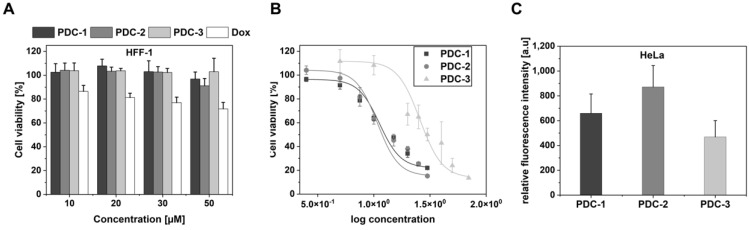
Herein, the design and synthesis of peptide-drug conjugates (PDCs) including different variants of the cell-penetrating peptide sC18 is presented. We first generated a series of novel sequence mutants of sC18 having either amino acid deletions and/or substitutions, and then tested their biological activity. The effects of histidine substituents were found to be not meaningful for sC18 uptake and cell selectivity. Moreover, building a nearly perfect amphipathic structure within a shortened sC18 derivative provided a peptide that was highly membrane-active, but also too cytotoxic. As a result, the most promising analog was sC18ΔE, which stands out due to its higher uptake efficacy compared to parent sC18. In the last set of experiments, we let the peptides react with the cytotoxic drug doxorubicin by Thiol-Michael addition to form novel PDCs. Our results indicate that sC18ΔE could be a more efficient drug carrier than parent sC18 for biomedical applications. However, cellular uptake using endocytosis and resulting entrapment of cargo inside vesicles is still a major critical step to overcome in CPP-containing peptide-drug development.

Keywords: cancer; cell-penetrating peptides; cytostatic drugs; drug delivery; peptide-drug conjugates.


Conflict of interest statement

The authors declare no conflict of interest.

Figures

Figure 1
Figure 1
(A,B): Cell-viability assays using MCF-7 cells. Cells were cultivated at pH 7.4 or pH 6.8, respectively, for 24 h using different concentrations of peptide solutions. Untreated cells served as negative control; cells treated with 70% ethanol as positive control. Values from the positive control were subtracted from all data, and the untreated cells were set to 100%; assays were conducted in triplicate n = 3. (C): Cellular uptake of histidine peptides in MCF-7 cells. Cells were cultivated at different pH-values and incubated with 10 µM peptide solutions for 30 min at 37 °C; assays were conducted in triplicate n = 3.
Figure 2
Figure 2
(A,B): Cytotoxicity profiles of the peptides in HeLa and HEK-293 cells. Cells were cultured and incubated at pH 7.4 for 24 h with different concentrations of peptide solutions. Untreated cells served as negative control; cells treated with 70% ethanol as positive control. Values from the positive control were subtracted from all data, and the untreated cells were set to 100%; assays were performed with n = 3 in triplicate. (C): Flow cytometry analysis of HeLa or HEK-293 cells after incubating 1 and 10 µM peptide solutions for 30 min at 37 °C; assays were conducted in triplicate n = 3. (D): Confocal laser scanning microscopy analysis of 10 µM CF-labeled peptides after 30 min of incubation in HeLa cells. Green: CF-labeled peptides; blue: Hoechst 33342 nuclear stain; scale bar is 10 µm.
Figure 3
Figure 3
(A): Flow cytometry analysis of HeLa spheroids that were incubated with 10 μM CF-labeled sC18 variants for 30 min and 1 h, respectively. (B): Flow cytometry analysis of HEK-293 spheroids that were incubated with 10 μM CF-labeled sC18 variants for 30 min and 1 h, respectively. (C): Distribution profiles for HeLa and HEK-293 spheroids after 30 min and 1 h incubation time. All experiments were conducted in triplicate with n = 4. (D): Proposed distribution profile of sC18-variants in tumor spheroids. Left: Accumulation of sC18-variants in HeLa spheroids. Right: Distribution of sC18-variants in HEK-293 spheroids [35].
Figure 4
Figure 4
Peptide interaction with giant lamellar vesicles (GUVs) composed of DOPC/DOPE/DOPG (40:30:30); 1 µM solutions of sC18 variants were incubated with GUVs for 30 min and inspected using a fluorescence microscope (Keyence). Red: Atto550; green: CF-labeled peptides, blue: Oyster 405. Scale bar: 50 µm.
Scheme 1
Scheme 1
Synthesis scheme of Dox-SMP-peptide conjugates. Exemplarily, the synthesis of PDC-2 is shown (see also Table 2 for peptide sequence).
Figure 5
Figure 5
(A): Cytotoxicity assay using HFF-1 cells. Different PDC concentrations were incubated with the cells for 24 h at 37 °C and a resazurin assay was conducted. (B): Dose-response curves for peptide drug conjugates were obtained using the resazurin assay. For this, HeLa cells were treated with various conjugate concentrations (2.5–70 µM) for 24 h at 37 °C. (C): Quantification of PDC internalization in HeLa cells; 10 µM PDC solutions were incubated with cells for 30 min at 37 °C and cells were inspected using a flow cytometer; assays were conducted in triplicate n = 3.
Figure 6
Figure 6
Fluorescence microscope analysis of 5 µM conjugates after 30 min of incubation at 37 °C in HeLa and HFF-1 cells. Red: peptides coupled to red fluorophore Doxorubicin; blue: Hoechst 33342 nuclear stain; BF: brightfield to identify living cells; scale bar is 20 µm.

References

    1. Derakhshankhah H., Jafari S. Cell penetrating peptides: A concise review with emphasis on biomedical applications. Biomed. Pharmacother. 2018;108:1090–1096. doi: 10.1016/j.biopha.2018.09.097. - DOI - PubMed
    1. Reissmann S. Cell penetration: Scope and limitations by the application of cell-penetrating peptides. J. Pept. Sci. 2014;20:760–784. doi: 10.1002/psc.2672. - DOI - PubMed
    1. Xie J., Bi Y., Zhang H., Dong S., Teng L., Lee R.J., Yang Z. Cell-Penetrating Peptides in Diagnosis and Treatment of Human Diseases: From Preclinical Research to Clinical Application. Front. Pharmacol. 2020;11:697. doi: 10.3389/fphar.2020.00697. - DOI - PMC - PubMed
    1. Gestin M., Dowaidar M., Langel Ü. Uptake mechanism of cell-penetrating peptides. Adv. Exp. Med. Biol. 2017;1030:255–264. - PubMed
    1. Sakamoto K., Akishiba M., Iwata T., Arafiles J.V.V., Imanishi M., Futaki S. Use of homoarginine to obtain attenuated cationic membrane lytic peptides. Bioorg. Med. Chem. Lett. 2021;40:127925. doi: 10.1016/j.bmcl.2021.127925. - DOI - PubMed

MeSH terms