. 2022 Oct 10;27(19):6747.
doi: 10.3390/molecules27196747.

Dextran Fluorescent Probes Containing Sulfadiazine and Rhodamine B Groups

Affiliations

Dextran Fluorescent Probes Containing Sulfadiazine and Rhodamine B Groups

Bi-Jie Bie et al. Molecules. .

Abstract

Fluorescent imaging has been expanded, as a non-invasive diagnostic modality for cancers, in recent years. Fluorescent probes in the near-infrared window can provide high sensitivity, resolution, and signal-to-noise ratio, without the use of ionizing radiation. Some fluorescent compounds with low molecular weight, such as rhodamine B (RhB) and indocyanine green (ICG), have been used in fluorescent imaging to improve imaging contrast and sensitivity; however, since these probes are excreted from the body quickly, they possess significant restrictions for imaging. To find a potential solution to this, this work investigated the synthesis and properties of novel macromolecular fluorescent compounds. Herein, water-soluble dextran fluorescent compounds (SD-Dextran-RhB) were prepared by the attachment of RhB and sulfadiazine (SD) derivatives to dextran carrier. These fluorescent compounds were then characterized through IR, 1H NMR, 13C NMR, UV, GPC, and other methods. Assays of their cellular uptake and cell cytotoxicity and fluorescent imaging were also performed. Through this study, it was found that SD-Dextran-RhB is sensitive to acidic conditions and possesses low cell cytotoxicities compared to normal 293 cells and HepG2 and HeLa tumor cells. Moreover, SD-Dextran-RhB demonstrated good fluorescent imaging in HepG2 and HeLa cells. Therefore, SD-Dextran-RhB is suitable to be potentially applied as a probe in the fluorescent imaging of tumors.

Keywords: dextran; fluorescent imaging; fluorescent probe; rhodamine B; sulfadiazine.


Conflict of interest statement

The authors declare no conflict of interest.

Figures

Scheme 1
Scheme 1
Synthetic route to dextran containing rhodamine B (Dextran-RhB).
Scheme 2
Scheme 2
Synthetic route to bromine-substituted dextran containing rhodamine B (Br-Dextran-RhB).
Scheme 3
Scheme 3
Synthetic route to dextran fluorescent compounds (SD-Dextran-RhB).
Figure 1
Figure 1
1H NMR and 13C NMR spectra of Dextran-RhB (a,b) and SD-Dextran-RhB (c,d).
Figure 1
Figure 1
1H NMR and 13C NMR spectra of Dextran-RhB (a,b) and SD-Dextran-RhB (c,d).
Figure 2
Figure 2
IR spectra (a) and DSC curves (b) of SD-Dextran-RhB.
Figure 3
Figure 3
GPC of dextran (a), Dextran-RhB (b), and SD-Dextran-RhB (c).
Figure 3
Figure 3
GPC of dextran (a), Dextran-RhB (b), and SD-Dextran-RhB (c).
Figure 4
Figure 4
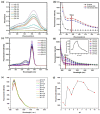
UV spectra (a), fluorescent spectra (b), fluorescent excitation spectra (c), and fluorescence emission spectra (d) of SD-Dextran-RhB and fluorescent excitation spectra (e,f) of RhB in different acidic conditions.
Figure 5
Figure 5
Cell toxicity assay of Dextran-RhB and SD-Dextran-RhB to normal 293 cells (a) and SD-Dextran-RhB (b) to HepG2 and HeLa cells.
Figure 5
Figure 5
Cell toxicity assay of Dextran-RhB and SD-Dextran-RhB to normal 293 cells (a) and SD-Dextran-RhB (b) to HepG2 and HeLa cells.
Figure 6
Figure 6
Confocal microscopy images (a) and relative intensity of fluorescent intensity (b) in HepG2 cells cultured with growth medium, Dextran-RhB, and SD-Dextran-RhB for 2 h.
Figure 7
Figure 7
Fluorescent imaging (a) and relative fluorescent intensity (b) of SD-Dextran-RhB in HeLa cells.

References

    1. Anderson C.J., Lewis J.S. Current status and future challenges for molecular imaging. Phil. Trans. R. Soc. A. 2017;375:20170023. doi: 10.1098/rsta.2017.0023. - DOI - PubMed
    1. Deng Y., Xu A., Yu Y., Fu C., Liang G. Biomedical applications of fluorescent and magnetic resonance imaging dual-modality probes. ChemBioChem. 2019;20:499–510. doi: 10.1002/cbic.201800450. - DOI - PubMed
    1. Zhang Y.B., Sun L., Yan Q., Qiu X.Y., Cheng Y.T., Wang B.L., Tan X.P., Fang M.X., Luck R.L., Liu H.Y. Near-infrared fluorescent probe based on cyanine scaffold for sensitive detection of uranyl ions in living cells and water samples. Microchem. J. 2022;180:107619. doi: 10.1016/j.microc.2022.107619. - DOI
    1. He S., Song J., Qu J., Cheng Z. Crucial breakthrough of second near-infrared biological window fluorophores: Design and synthesis toward multimodal imaging and theranostics. Chem. Soc. Rev. 2018;47:4258–4278. doi: 10.1039/C8CS00234G. - DOI - PubMed
    1. Huang X., Song J., Yung B.C., Huang X., Xiong Y., Chen X. Ratiometric optical nanoprobes e;nable accurate molecular detection and imaging. Chem. Soc. Rev. 2018;47:2873–2920. doi: 10.1039/C7CS00612H. - DOI - PMC - PubMed

LinkOut - more resources