. 2022 Sep 22;14(19):3938.
doi: 10.3390/nu14193938.

Ginsenosides Restore Lipid and Redox Homeostasis in Mice with Intrahepatic Cholestasis through SIRT1/AMPK Pathways

Affiliations

Ginsenosides Restore Lipid and Redox Homeostasis in Mice with Intrahepatic Cholestasis through SIRT1/AMPK Pathways

Guodong Li et al. Nutrients. .

Abstract

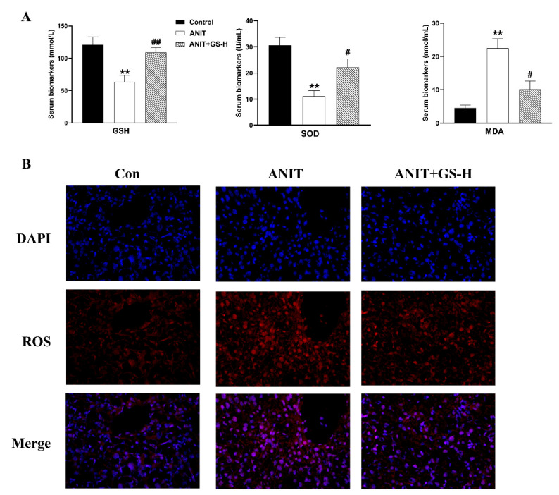
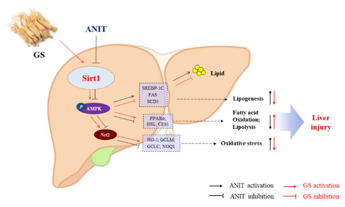
Intrahepatic cholestasis (IC) occurs when the liver and systemic circulation accumulate bile components, which can then lead to lipid metabolism disorders and oxidative damage. Ginsenosides (GS) are pharmacologically active plant products derived from ginseng that possesses lipid-regulation and antioxidation activities. The purpose of this study was to evaluate the possible protective effects of ginsenosides (GS) on lipid homeostasis disorder and oxidative stress in mice with alpha-naphthylisothiocyanate (ANIT)-induced IC and to investigate the underlying mechanisms. A comprehensive strategy via incorporating pharmacodynamics and molecular biology technology was adopted to investigate the therapeutic mechanisms of GS in ANIT-induced mice liver injury. The effects of GS on cholestasis were studied in mice that had been exposed to ANIT-induced cholestasis. The human HepG2 cell line was then used in vitro to investigate the molecular mechanisms by which GS might improve IC. The gene silencing experiment and liver-specific sirtuin-1 (SIRT1) knockout (SIRT1LKO) mice were used to further elucidate the mechanisms. The general physical indicators were assessed, and biological samples were collected for serum biochemical indexes, lipid metabolism, and oxidative stress-related indicators. Quantitative PCR and H&E staining were used for molecular and pathological analysis. The altered expression levels of key pathway proteins (Sirt1, p-AMPK, Nrf2) were validated by Western blotting. By modulating the AMPK protein expression, GS decreased hepatic lipogenesis, and increased fatty acid β-oxidation and lipoprotein lipolysis, thereby improving lipid homeostasis in IC mice. Furthermore, GS reduced ANIT-triggered oxidative damage by enhancing Nrf2 and its downstream target levels. Notably, the protective results of GS were eliminated by SIRT1 shRNA in vitro and SIRT1LKO mice in vivo. GS can restore the balance of the lipid metabolism and redox in the livers of ANIT-induced IC models via the SIRT1/AMPK signaling pathway, thus exerting a protective effect against ANIT-induced cholestatic liver injury.

Keywords: antioxidant; ginsenosides (GS); intrahepatic cholestasis (IC); lipid metabolism.


Conflict of interest statement

The authors declared no potential conflict of interest with respect to the research, authorship, and/or publication of this article. The entire thesis or any part of manuscript has not been published or accepted elsewhere. It has not been submitted to any other journals.

Figures

Figure 1
Figure 1
Structural formula of the ginsenoside (GS) monomer. The numerical superscript indicates the carbon on the glycosidic bond. Glc, glucose; Ara(f), arabinofuranose; Rha, rhamnose.
Figure 2
Figure 2
GS markedly improved cholestatic liver injury in ANIT-induced mice. (A) Body weight changes of mice from day 1–5. (B) Comparison of liver weight in each group on the fifth day. (C) Comparison of liver weight/body weight ratio in each group on the fifth day. (D) Histopathology in hematoxylin and eosin-stained liver sections (Scale bar, 50 um). Black arrows, histological damage. (E,F) The levels of ALT, AST, ALP, TBA, TBIL, and DBIL in serum of mice. Significantly different from control, ** p < 0.01, or ANIT, # p < 0.05 or ## p < 0.01.
Figure 3
Figure 3
GS treatment improved expression of genes involved in bile acid homeostasis in the liver. (A,B) The mRNA levels and protein levels of bile metabolism-related genes (CYP7A1, CYP27A1, NTCP, BSEP, MRP2) in mice after ANIT and GS exposure. Significantly different from control, * p < 0.05 or ** p < 0.01, or ANIT, # p < 0.05.
Figure 4
Figure 4
GS treatment improved fat metabolism gene profile in murine liver. (A) Morphology of frozen liver sections as tested by Oil Red O staining (scale bar, 20 μm). Red arrows, lipid droplets. (B) Levels of TG, TC, HDL, and LDL in serum of mice. (C,D) The levels of mRNAs (ACC2, PPARα, SREBP-1, FAS, SCD1, ACC1, ChREBP, HMGCR) and proteins (PPARα, SREBP-1, FAS, SCD1, HSL, CES1) of fat metabolism-related genes in mice after ANIT and GS exposure. Significantly different from control, * p < 0.05 or ** p < 0.01, or ANIT, # p < 0.05 or ## p < 0.01.
Figure 5
Figure 5
GS treatment improved oxidative damage in ANIT-induced mice. (A) The levels of GSH, SOD, and MDA in serum of mice. (B) Immunofluorescence stainings for ROS levels in liver tissue. Significantly different from control, ** p < 0.01, or ANIT, # p < 0.05 or ## p < 0.01.
Figure 6
Figure 6
GS treatment improved the expression level of oxidative stress genes. (A,B) The mRNA and protein expressions of genes related to induce oxidative stress or associated genes (Nrf2, HO-1, GCLM, GCLC, and NQO1). Significantly different from control, * p < 0.05 or ** p < 0.01, or ANIT, # p < 0.05 or ## p < 0.01.
Figure 7
Figure 7
GS treatment improved the protein expression level of SIRT1/AMPK. (A) The protein expression levels of SIRT1, p-AMPK, and AMPK. (B) Immunofluorescence stainings for SIRT1 and p-AMPK protein levels in liver tissue. Significantly different from control, * p < 0.05 or ** p < 0.01, or ANIT, # p < 0.05 or ## p < 0.01.
Figure 8
Figure 8
GS activated the SIRT1/AMPK signaling pathway in HepG2 cells. (A,B) Levels of TG, TC, GSH, and SOD in cell extracts. (C) Immunofluorescent analysis of neutral lipids accumulation (fat droplets) in HepG2 cells using BODIPY stain. (D) The production of ROS in HepG2 cells was assessed by DCHF-DA assay. (E,F) After the addition of sh-SIRT1 or doxorubicin hydrochloride (DH, AMPK inhibitor) the role of GS on SIRT1, p-AMPK, AMPK, and Nrf2 levels was tested by Western blot. Significantly different from control, * p < 0.05 or ** p < 0.01, or ANIT, # p < 0.05 or ## p < 0.01, or ANIT+GS-H, & p < 0.05 or && p < 0.01.
Figure 9
Figure 9
The therapeutic effect of GS was absent in SIRT1LKO mice. (A) Liver weight changes (WT compared with SIRT1LKO. (BF) The biochemical indexes of ALT, AST, TBA, TG, TC, GSH, and SOD in the serum of mice. (G) Histopathology in hematoxylin and eosin-stained liver sections (scale bar, 50 µm). Black arrows, histological damage. Morphology of frozen liver sections as tested by Oil Red O staining (scale bar, 20 µm). Red arrows, lipid droplets. Significantly different from ANIT (WT), * p < 0.05 or ** p < 0.01, or ANIT+GS (WT), # p < 0.05 or ## p < 0.01.
Figure 10
Figure 10
GS-induced activation of the SIRT1/AMPK signaling pathway was significantly reversed in SIRT1LKO mice. (A) Immunofluorescent staining for ROS levels in liver tissue. (B) Protein levels of SIRT1, p-AMPK, AMPK, and Nrf2 levels in WT and SIRT1LKO after GS treatment. Significantly different from ANIT (WT), ** p < 0.01, or ANIT+GS (WT), ## p < 0.01.
Figure 11
Figure 11
The proposed mechanism of GS-mediated protection in IC via the activation of the SIRT2/AMPK pathway. GS increases expression of SIRT1, leading to phosphorylation of AMPK and Nrf2, and the subsequent modulation of genes associated with inhibiting hepatic lipogenesis and increasing hepatic fatty acid oxidation and lipolysis.

References

    1. Dyson J.K., Hirschfield G.M., Adams D.H., Beuers U., Mann D.A., Lindor K.D., Jones D.E.J. Novel therapeutic targets in primary biliary cirrhosis. Nat. Rev. Gastro. Hepat. 2015;12:147–158. doi: 10.1038/nrgastro.2015.12. - DOI - PubMed
    1. Yu L.X., Liu X.X., Yuan Z.H., Li X.J.Y., Yang H., Yuan Z.Q., Sun L.X., Zhang L.Y., Jiang Z.Z. SRT1720 Alleviates ANIT-Induced Cholestasis in a Mouse Model. Front. Pharmacol. 2017;8:256. doi: 10.3389/fphar.2017.00256. - DOI - PMC - PubMed
    1. Manos M.M., Leyden W.A., Murphy R.C., Terrault N.A., Bell B.P. Limitations of conventionally derived chronic liver disease mortality rates: Results of a comprehensive assessment. Hepatology. 2008;47:1150–1157. doi: 10.1002/hep.22181. - DOI - PubMed
    1. Menżyk T., Bator M., Derra A., Kierach R., Kukla M. The role of metabolic disorders in the pathogenesis of intrahepatic cholestasis of pregnancy. Clin. Exp. Hepatol. 2018;4:217–223. doi: 10.5114/ceh.2018.80122. - DOI - PMC - PubMed
    1. Dann A.T., Kenyon A.P., Wierzbicki A.S., Seed P.T., Shennan A.H., Tribe R.M. Plasma Lipid Profiles of Women with intrahepatic cholestasis of pregnancy. Obstet. Gynecol. 2006;107:106–114. doi: 10.1097/01.AOG.0000189096.94874.9c. - DOI - PubMed

MeSH terms

LinkOut - more resources