Review
. 2022 Sep 28;22(19):7374.
doi: 10.3390/s22197374.

Development and Applications of Compton Camera-A Review

Affiliations
Review

Development and Applications of Compton Camera-A Review

Raj Kumar Parajuli et al. Sensors (Basel). .

Abstract

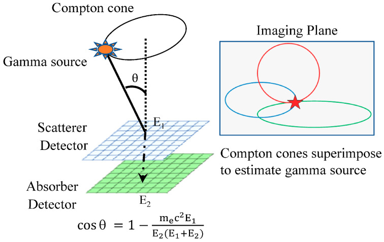
The history of Compton cameras began with the detection of radiation sources originally for applications in astronomy. A Compton camera is a promising γ-ray detector that operates in the wide energy range of a few tens of keV to MeV. The γ-ray detection method of a Compton camera is based on Compton scattering kinematics, which is used to determine the direction and energy of the γ-rays without using a mechanical collimator. Although the Compton camera was originally designed for astrophysical applications, it was later applied in medical imaging as well. Moreover, its application in environmental radiation measurements is also under study. Although a few review papers regarding Compton cameras have been published, they either focus very specifically on the detectors used in such cameras or the particular applications of Compton cameras. Thus, the aim of this paper is to review the features and types of Compton cameras and introduce their applications, associated imaging algorithms, improvement scopes, and their future aspects.

Keywords: Compton camera; detectors; medical imaging; γ-rays.


Conflict of interest statement

The authors declare no conflict of interest.

Figures

Figure 1
Figure 1
Energy at which photoelectric effect, Compton scattering, and pair production are dominant with respect to atomic number (Z) of the absorber.
Figure 2
Figure 2
Schematic illustration of a general Compton camera (left); Compton cones of each event are superimposed to locate the γ-ray source (right).
Figure 3
Figure 3
Photographs of (a) multilayered DSSDs stack [65], (b) the CdTe detectors stack [65], and (c) the prototype Si/CdTe Compton camera [66]. Reproduced with permissions from Watanabe et al. [65] and Takeda et al. [66].
Figure 4
Figure 4
Pixelated HPGe Compton camera with the detector configurations [109]. Reprinted with permissions from Alnaaimi et al. [109].
Figure 5
Figure 5
Ce:GAGG and MPPC based DOI handy Compton camera developed by Kataoka et al. [118]. Left photograph shows the Compton camera in compact form and the right picture shows the internal structure of the camera. Reprinted with permissions from Kataoka et al. [118].
Figure 6
Figure 6
Conceptual structure (left (a) ETCC (b) tracks of electrons and (c) schematic structure of micro-pixel gas chamber) and actual photograph of ETCC (right) developed by Kabuki et al. [129]. Reproduced with permissions from Kabuki et al. [129].
Figure 7
Figure 7
1–30 MeV gamma in the sky as observed by COMPTEL in the 1990s (upper left) and the simulated Cygnus region in the 1–30 MeV energy region expected from e-ASTROGAM (lower right) [151]. Reproduced with permissions from Angelis et al. [151].
Figure 8
Figure 8
(a) Human experiment setup using Compton camera. (b) Energy spectrum obtained in the study. (c) Compton image overlaid with CT image for 99mTc DMSA radiopharmaceuticals. (d) Compton image overlaid with CT image for 18F FDG radiopharmaceuticals. All the images are reprinted from Nakano et al. [84]. Reprinted with permissions from Nakano et al. [84].
Figure 9
Figure 9
In vivo Compton imaging of accumulation of (a) 111I in mouse tumor and liver, (b) 18F-FDG in heart and bladder, and (c) The PET imaging of 18F-FDG accumulated in mouse heart. The images were superimposed with the CT images [156]. Reproduced with permissions from Uenomachi et al. [156].
Figure 10
Figure 10
(a) Configuration of multi-angle data acquisition measurement of Ce:GAGG Compton camera. (b) Energy spectrum obtained via 10-min measurements from an angle in a mouse injected with 131I, 85Sr, and 65Zn. (c) Compton image of 131I, (d) 85Sr, and (e) 65Zn, and (f) fused images of all three tracers. All the figures are reprinted from Kishimoto et al. [155]. Reproduced with permissions from Kishimoto et al. [155].
Figure 11
Figure 11
In vivo real-time monitoring of annihilation γ-rays generated by CIRT using Si/CdTe Compton camera. Experimental setup (left). Compton image of 511 keV annihilation gammas (right). Images have been rearranged and reprinted from Shiba et al. [87].
Figure 12
Figure 12
On-beam range monitoring using Si/CdTe Compton camera in CIRT. (a) Experimental setup. (b) Compton image of 511 keV annihilation gammas [85]. (c) Compton image of 718 keV prompt gammas [86]. Dotted line in figures (b,c) represents the phantom periphery. All the figures are rearranged and reprinted from Parajuli et al. [85,86].
Figure 13
Figure 13
(a) Drone used for carrying Compton camera for radiation monitoring. (b) Zoomed view of radiation measurement system. (c) Optical image taken using drone at altitude of 6 m. (d) Radiation image measured using the drone system in which the a, b, and c regions represent the hotspots [181]. Reproduced with permissions from Sato et al. [181].
Figure 14
Figure 14
Example of an implication of event position by the SOE method. The event may move to the new location or remain at the current (old) location, according to the acceptance probability equation: (a) a new position will be accepted with 100% probability as in this case the event density increases; (b) the new position will be accepted with low probability due to the sharp decrease in event density. The figures are rearranged and reprinted with permissions from Andreyev et al. [218].

Similar articles

Cited by

References

    1. Schoenfelder V., Hirner A., Schneider K. A telescope for soft gamma astronomy. Nucl. Instrum. Methods. 1973;107:385–394. doi: 10.1016/0029-554X(73)90257-7. - DOI
    1. Herzo D., Koga R., Millard W.A., Moon S., Ryan J., Wilson R., Zych A.D., White R.S. A large double scatter telescope for gamma rays and neutrons. Nucl. Instrum. Methods. 1975;123:583. doi: 10.1016/0029-554X(75)90215-3. - DOI
    1. Lockwood J.A., Hsieh L., Friling L., Chen C., Swartz D. Atmospheric neutron and gamma ray fluxes and energy spectra. J. Geophys. Res. 1979;84:1402. doi: 10.1029/JA084iA04p01402. - DOI
    1. Schoenfelder V., Aarts H., Bennett K., de Boer H., Clear J., Collmar W., Connors A., Deerenberg A., Diehl R., von Dordrecht A., et al. Instrument description and performance of the imaging gamma-ray telescope COMPTEL aboard the Compton gamma-ray observatory. Astrophys. J. 1993;86:657. doi: 10.1086/191794. - DOI
    1. Todd R., Nightingale J.M., Everett D.B. A proposed γ camera. Nature. 1974;251:132–134. doi: 10.1038/251132a0. - DOI