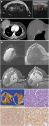
Extraskeletal Ewing Sarcoma of the Chest Wall Manifesting as a Palpable Breast Mass: Ultrasonography, CT, and MRI Findings
- PMID: 36237473
- PMCID: PMC9432414
- DOI: 10.3348/jksr.2020.0109
Extraskeletal Ewing Sarcoma of the Chest Wall Manifesting as a Palpable Breast Mass: Ultrasonography, CT, and MRI Findings
Abstract
Ewing sarcomas constitute a group of small, round, blue cell tumors of the bone and soft tissue. Extraskeletal Ewing sarcoma (EES) is a rare malignant neoplasm that arises from soft tissues, and it usually affects children and young adults. EES of the thoracopulmonary region commonly presents with a palpable mass or pain. Although rarely reported, EES affecting the anterior chest wall may present as a breast mass. We report a case of EES arising from the chest wall and manifesting as a palpable breast mass in a 22-year-old woman. The large mass was initially misdiagnosed as a breast origin mass on ultrasonography, but subsequent CT and MRI showed that the mass originated from the chest wall. Radiologists should be aware of the imaging findings of EES, and they should understand that chest wall lesions may be clinically confused as breast lesions.
유잉육종계열의 종양은 뼈와 연부조직에 발생하는 악성 소원형청색세포종양이다. 골격외 유잉씨 육종은 드문 악성 종양으로 연부조직에 발생한 유잉육종의 한 형태이며, 소아와 젊은 성인에서 호발한다. 흉폐부위에 발생한 골격외 유잉씨 육종은 임상적으로 만져지는 종괴나 통증으로 나타난다. 골격외 유잉씨 육종이 앞가슴벽을 침범한 경우에는 유방 종괴로 나타날 수 있으나, 이러한 보고는 드물다. 저자들은 22세 여성에서 유방 종괴로 나타난 앞가슴벽에 발생한 유잉씨 육종의 증례를 보고한다. 초기의 초음파에서 이 거대 종괴는 유방에서 발생한 종괴로 오인되었으나, 추가적인 전산화단층촬영 및 자기공명영상에서 종괴는 흉벽에서 기원하였음을 알 수 있었다. 영상의학과 의사는 골격외 유잉씨 육종의 영상 소견을 알고, 흉벽의 병변이 임상적으로 유방 병변으로 오인될 수 있음을 이해하는 것이 중요하다.
Keywords: Chest Wall; Computed Tomography, X-Ray; Ewing Sarcoma; Magnetic Resonance Imaging; Ultrasonography.
Copyrights © 2021 The Korean Society of Radiology.
Conflict of interest statement
Conflicts of Interest: The authors have no potential conflicts of interest to disclose.
Figures

Similar articles
-
Case report: Primary pleural giant extraskeletal Ewing sarcoma in a child.Front Oncol. 2023 Mar 31;13:1137586. doi: 10.3389/fonc.2023.1137586. eCollection 2023. Front Oncol. 2023. PMID: 37064103 Free PMC article.
-
A case of extraskeletal Ewing sarcoma originating from the visceral pleura.Hippokratia. 2011 Oct;15(4):363-5. Hippokratia. 2011. PMID: 24391423 Free PMC article.
-
Imaging-based diagnosis for extraskeletal Ewing sarcoma in pediatrics: A case report.World J Clin Cases. 2022 Jul 6;10(19):6595-6601. doi: 10.12998/wjcc.v10.i19.6595. World J Clin Cases. 2022. PMID: 35979293 Free PMC article.
-
From the radiologic pathology archives: ewing sarcoma family of tumors: radiologic-pathologic correlation.Radiographics. 2013 May;33(3):803-31. doi: 10.1148/rg.333135005. Radiographics. 2013. PMID: 23674776 Review.
-
Extraskeletal Ewing Sarcoma from Head to Toe: Multimodality Imaging Review.Radiographics. 2022 Jul-Aug;42(4):1145-1160. doi: 10.1148/rg.210226. Epub 2022 May 27. Radiographics. 2022. PMID: 35622491 Review.
References
-
- Murphey MD, Senchak LT, Mambalam PK, Logie CI, Klassen-Fischer MK, Kransdorf MJ. From the radiologic pathology archives: ewing sarcoma family of tumors: radiologic-pathologic correlation. Radiographics. 2013;33:803–831. - PubMed
-
- Javery O, Krajewski K, O'Regan K, Kis B, Giardino A, Jagannathan J, et al. A to Z of extraskeletal Ewing sarcoma family of tumors in adults: imaging features of primary disease, metastatic patterns, and treatment responses. AJR Am J Roentgenol. 2011;197:W1015–W1022. - PubMed
-
- Choi YC, Oh YW, Park CM, Chung KB, Choi MS, Choi YH. Malignant small cell tumor of the thoracopulmonary region (Askin tumor) J Korean Radiol Soc. 1989;25:47–51.
-
- Hari S, Jain TP, Thulkar S, Bakhshi S. Imaging features of peripheral primitive neuroectodermal tumours. Br J Radiol. 2008;81:975–983. - PubMed
-
- Carter BW, Benveniste MF, Betancourt SL, De Groot PM, Lichtenberger JP, 3rd, Amini B, et al. Imaging evaluation of malignant chest wall neoplasms. Radiographics. 2016;36:1285–1306. - PubMed
Publication types
LinkOut - more resources
Full Text Sources

