Review
. 2021 Jul;82(4):826-837.
doi: 10.3348/jksr.2021.0078. Epub 2021 Jul 26.

[Immunoglobulin G4-Related Disease in the Thorax: Imaging Findings and Differential Diagnosis]

[Article in Korean]
Review

[Immunoglobulin G4-Related Disease in the Thorax: Imaging Findings and Differential Diagnosis]

[Article in Korean]
Yookyung Kim et al. Taehan Yongsang Uihakhoe Chi. 2021 Jul.

Abstract

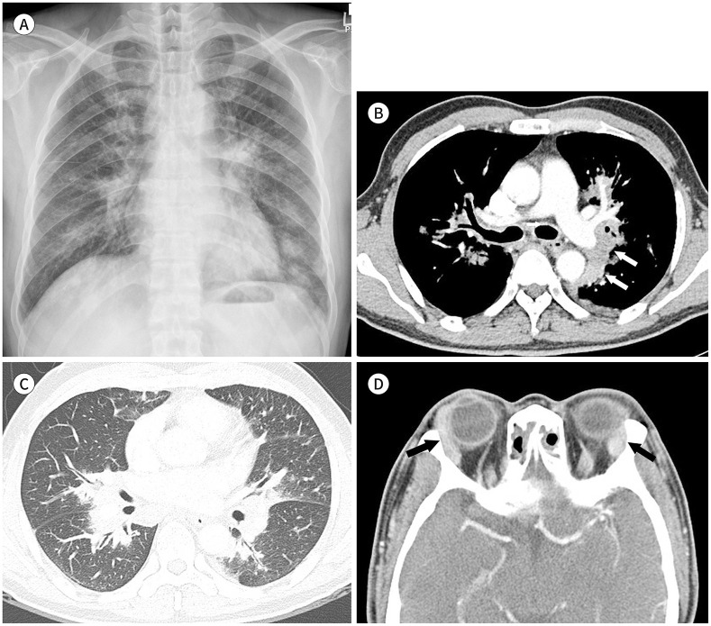
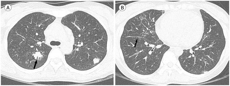
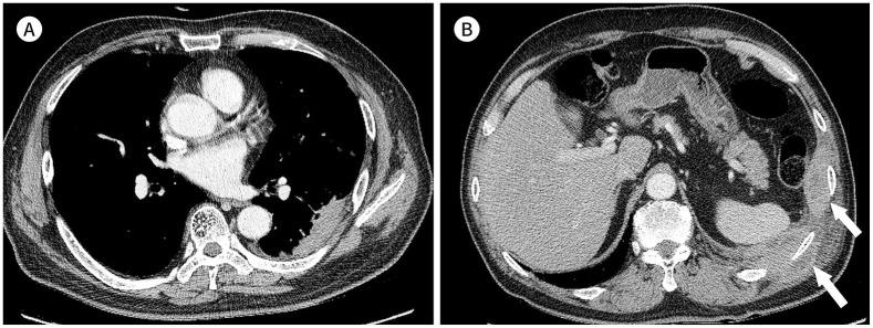
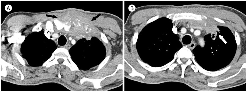
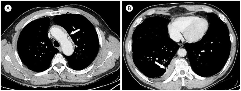
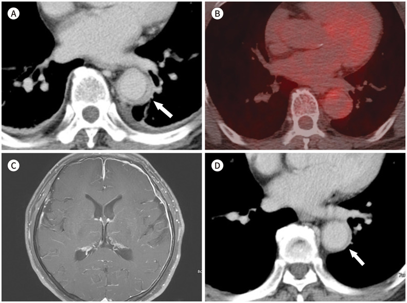
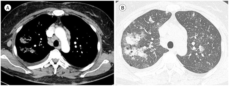
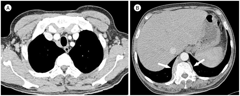
Immunoglobulin G4-related disease (IgG4-RD) is a chronic inflammatory condition involving multiple organs, including the salivary or lacrimal glands, orbit, pancreas, bile duct, liver, kidney, retroperitoneum, aorta, lung, and lymph nodes. It is histologically characterized by tissue infiltration with lymphocytes and IgG4-secreting plasma cells, storiform fibrosis, and obliterative phlebitis. In the thoracic involvement of IgG4-RD, mediastinal lymphadenopathy and perilymphangitic interstitial thickening of the lung are the most common findings. Peribronchovascular and septal thickening and paravertebral band-like soft tissue are characteristic findings of IgG4-RD. Other findings include pulmonary nodules or masses, ground-glass opacity, alveolar interstitial thickening, pleural effusion or thickening, mass in the chest wall or mediastinum, and arteritis involving the aorta and coronary artery. Radiologic differential diagnosis of various malignancies, infections, and inflammatory conditions is needed. In this review, we describe the imaging findings of IgG4-RD and the radiologic differential diagnoses in the thorax.

면역글로불린G4 연관 질환(immunoglobulin G4-related disease; 이하 IgG4-RD)는 IgG4를 생산하는 면역세포에 의한 만성 염증성질환으로, 주로 타액선, 누액선, 안와, 췌장, 담도, 간, 신장, 후복막, 대동맥, 폐, 림프절 등 다양한 장기를 침범하고, 조직학적으로 IgG4 양성 형질세포와 림프구의 침윤 및 나선형의 섬유화(storiform fibrosis), 폐색정맥염(obliterative phlebitis)을 특징으로 한다. IgG4-RD의 흉부 침범에서 가장 흔한 소견은 종격동 림프절 비대와 폐의 림프관주위 간질 비후이다. 폐의 기관지혈관주위 간질 비후와 우측 척추곁 밴드형 연부조직은 IgG4-RD의 특징적 소견이고, 그 외에도 폐결절 혹은 종괴, 간유리음영, 폐포 간질비후, 흉막삼출 및 비후, 흉벽이나 종격동 종괴, 대동맥과 관상동맥의 혈관염이 발생할 수 있다. 영상의학적으로는 악성 종양이나 감염 및 다양한 염증성질환과의 감별진단이 필요하다. 본 연구에서는 흉부에서 발생하는 IgG4-RD의 영상 소견과 감별진단에 대해 기술하였다.


Conflict of interest statement

Conflicts of Interest: The authors have no potential conflicts of interest to disclose.

Figures

Fig. 1
Fig. 1. A 56-year-old male with immunoglobulin G4-related disease involving the lung, lymph nodes, and lacrimal glands.
A. Chest radiograph shows perihilar and peribronchial infiltrations and poorly defined nodules in both lungs. B, C. CT images show diffuse peribrochovascular interstitial thickening, interlobular septal thickening, and ground-glass opacities in both lungs. In addition, mediastinal and bilateral hilar lymphadenopathy and mediastinal soft tissue mass are shown around the descending thoracic aorta and pulmonary artery (B) (arrows). D. Contrast-enhanced brain CT demonstrates enlargements of both lacrimal glands (arrows).
Fig. 2
Fig. 2. A 39-year-old female with immunoglobulin G4-related disease involving the lungs and lymph nodes.
A. Axial CT scan shows poorly defined nodules in both upper lobes. The nodule in the right upper lobe reveals an air bronchogram (arrow) and ground-glass opacity in the peripheral portion. B. Interlobular septal and peribronchovasuclar interstitial thickenings and patchy areas of ground-glass opacities are noted in the right middle lobe, associated with nodular thickening of the right major fissure (arrow).
Fig. 3
Fig. 3. A 67-year-old male with immunoglobulin G4-related disease involving the lung and chest wall.
A, B. Contrast-enhanced CT images show an ill-defined mass-like lesion in the left lower lobe (A) and two soft-tissue masses showing areas of low attenuation in the left chest wall (B) (arrows). Courtesy Yoon Kyung Kim, MD, Samsung Medical Center.
Fig. 4
Fig. 4. A 64-year-old male with immunoglobulin G4-related disease presenting with a chest wall mass at the sternoclavicular joint.
A, B. Contrast-enhanced CT images demonstrate ill-defined mas-like soft tissue infiltration in the left anterior chest wall at the sternoclavicular joint (black arrows). Bony destruction in the chest wall mass (A) and infiltration into the adjacent left upper lobe (B) (white arrow) are noted.
Fig. 5
Fig. 5. Immunoglobulin G4-related aortitis and periaortitis, and paravertebral soft tissue in a 65-year-old male.
A. Axial contrast-enhanced delayed-phase CT image showing diffuse wall thickening with homogeneous enhancement at the aortic arch (arrow). B. A band-like mass of soft tissue is noted at the right paravertebral area (arrow). Courtesy of Yoon Kyung Kim, MD, Samsung Medical Center.
Fig. 6
Fig. 6. Immunoglobulin G4-related thoracic aortitis and pachymeningitis in a 64-year-old male.
A, B. Axial contrast-enhanced delayed phase CT image showing homogeneously enhanced wall thickening of the descending thoracic aorta (A) (arrow), which demonstrates focal FDG uptake on axial FDG PET/CT images (maximal standard uptake value, 4.5), indicative of active inflammation (B). C. Gadolinium-enhanced brain MR image reveals diffuse and nodular-enhancing thickening of the falx cerebri and bilateral cerebral convexities. D. Aortic wall thickening has been resolved on follow-up CT scan obtained after steroid treatment (arrow). Courtesy of Eun Ju Chun, MD, Seoul National University Bundang Hospital. FDG = fluorodeoxyglucose
Fig. 7
Fig. 7. Immunoglobulin G4-related coronary arteritis and periarteritis in a 60-year-old male.
A–C. Echocardiography-gated coronary CT angiography images demonstrate multifocal segmental wall thickening with significant luminal narrowing at the left anterior descending (A, B) and right coronary arteries (C) (arrows). Courtesy of Eun Ju Chun, MD, Seoul National University Bundang Hospital.
Fig. 8
Fig. 8. T-cell lymphoma in a 52-year-old female.
A. Contrast-enhanced CT scan shows mediastinal and right axillary lymphadenopathy. B. Lung window image reveals peribronchial and interlobular septal thickenings, areas of ground-glass opacity and consolidation, and poorly defined nodules in both lungs.
Fig. 9
Fig. 9. A 45-year-old male with multicentric Castleman's disease.
A. Axial CT scan shows mediastinal lymphadenopathy. B. Diffuse perilymphatic pulmonary interstitial thickenings, including peribronchial and interlobular septal thickenings, left major fissural thickening, and ground-glass opacity, are observed in both lungs.
Fig. 10
Fig. 10. A 64-year-old female with Erdheim-Chester disease.
A, B. Contrast-enhanced axial CT scans demonstrate periaortic soft tissue around the aortic arch and descending thoracic aorta (arrows). C. Lung window image reveals diffuse peribronchial and interlobular septal thickening and thickened major fissures. D. Technetium 99m-methyl diphosphonate bone scan shows bilateral symmetric increased uptake in the meta-diaphyseal region in the distal femurs and proximal and distal tibiae. In addition, increased uptake is seen in the distal radii, maxilla, mandible, L4 vertebra, and right pubic bone.
Fig. 11
Fig. 11. A 77-year-old male with lymphoplasmacytic lymphoma.
A. Contrast-enhanced axial CT scan shows enlarged lymph nodes in the mediastinum and both axillae. B. A paravertebral band-like mass of soft tissue is noted, predominantly in the right paravertebral area (arrows).
Fig. 12
Fig. 12. Three cases of chest wall infection caused by different organisms.
A–C. A soft tissue mass involving the chest wall is noted in cases of actinomycosis (A), tuberculosis (B), and acute suppurative bacterial infection (C) (arrows).

Similar articles

  • Immunoglobulin G4-related hepatobiliary disease.
    Lee HE, Zhang L. Lee HE, et al. Semin Diagn Pathol. 2019 Nov;36(6):423-433. doi: 10.1053/j.semdp.2019.07.007. Epub 2019 Jul 24. Semin Diagn Pathol. 2019. PMID: 31358425 Review.
  • Intrathoracic Involvements of Immunoglobulin G4-Related Sclerosing Disease.
    Fei Y, Shi J, Lin W, Chen Y, Feng R, Wu Q, Gao X, Xu W, Zhang W, Zhang X, Zhao Y, Zeng X, Zhang F. Fei Y, et al. Medicine (Baltimore). 2015 Dec;94(50):e2150. doi: 10.1097/MD.0000000000002150. Medicine (Baltimore). 2015. PMID: 26683924 Free PMC article.
  • Immunoglobulin G4-Related Disease: What an Allergist Should Know.
    Carballo I, González-Quintela A, Sopeña B, Vidal C. Carballo I, et al. J Investig Allergol Clin Immunol. 2021 Jun 22;31(3):212-227. doi: 10.18176/jiaci.0633. Epub 2020 Jul 30. J Investig Allergol Clin Immunol. 2021. PMID: 32732179 Review.
  • Thoracic manifestations of IgG4-related disease.
    Muller R, Ebbo M, Habert P, Daniel L, Briantais A, Chanez P, Gaubert JY, Schleinitz N. Muller R, et al. Respirology. 2023 Feb;28(2):120-131. doi: 10.1111/resp.14422. Epub 2022 Nov 27. Respirology. 2023. PMID: 36437514 Free PMC article. Review.
  • Thoracic involvement and imaging patterns in IgG4-related disease.
    Muller R, Habert P, Ebbo M, Graveleau J, Groh M, Launay D, Audia S, Pugnet G, Cohen F, Perlat A, Benyamine A, Bienvenu B, Gaigne L, Chanez P, Gaubert JY, Schleinitz N. Muller R, et al. Eur Respir Rev. 2021 Oct 5;30(162):210078. doi: 10.1183/16000617.0078-2021. Print 2021 Dec 31. Eur Respir Rev. 2021. PMID: 34615698 Free PMC article. Review.

Cited by

References

    1. Kamisawa T, Zen Y, Pillai S, Stone JH. IgG4-related disease. Lancet. 2015;385:1460–1471. - PubMed
    1. Stone JH, Zen Y, Deshpande V. IgG4-related disease. N Engl J Med. 2012;366:539–551. - PubMed
    1. Corcoran JP, Culver EL, Anstey RM, Talwar A, Manganis CD, Cargill TN, et al. Thoracic involvement in IgG4-related disease in a UK-based patient cohort. Respir Med. 2017;132:117–121. - PubMed
    1. Zhang Z, Guan W, Lin Q, Yu W. Thoracic paravertebral involvement in patients with IgG4-related disease: CT and MR imaging findings. Rheumatology (Oxford) 2020;59:3878–3885. - PubMed
    1. Kanno A, Nishimori I, Masamune A, Kikuta K, Hirota M, Kuriyama S, et al. Nationwide epidemiological survey of autoimmune pancreatitis in Japan. Pancreas. 2012;41:835–839. - PubMed