

Beaver tail liver on pediatric chest X-ray
- PMID: 36238209
- PMCID: PMC9550850
- DOI: 10.1016/j.radcr.2022.09.025
Beaver tail liver on pediatric chest X-ray
Abstract
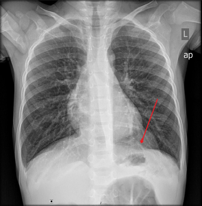
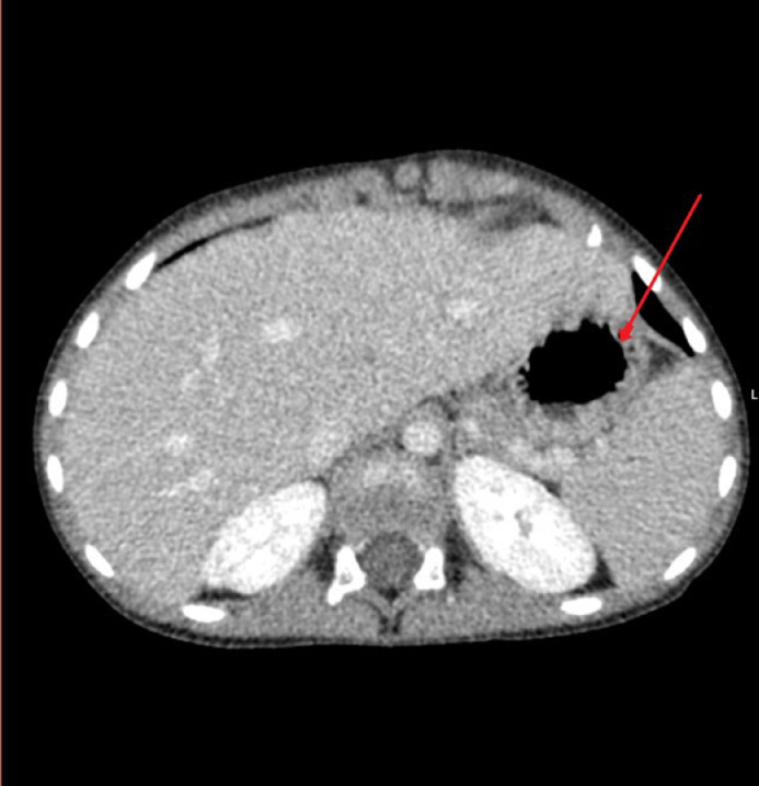
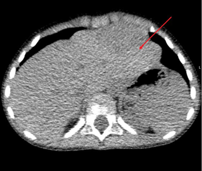
Beaver tail liver is an anatomical liver variant presenting as elongated left lobe of liver which extends laterally to the spleen. It can present with symptoms or be detected accidentally. We present a case of a 2-year-old asymptomatic patient who was had an X-ray of the chest describing a shadow of unknown origin located left paracardial and the diagnosis of "beaver tail liver" was confirmed after a multi-slice computed tomography of thorax and abdomen was done. We give an overview of very sparse available literature on this anatomical variant while emphasizing the rarity of diagnosis in pediatric population and clinical importance of this variant.
Keywords: Beaver tail liver; Liver; MSCT; Pediatric; X-ray.
© 2022 The Authors. Published by Elsevier Inc. on behalf of University of Washington.
Figures
References
-
- Atalar MH, Karakus K. Beaver tail liver. Abdom Radiol (NY) 2018;43:1851–1852. - PubMed
-
- Xiang H, Han J, Ridley WE, Ridley LJ. Beaver tail liver: anatomic variant. J Med Imaging Radiat Oncol. 2018;62:57. - PubMed
-
- Crivello MS, Peterson IM, Austin RM. Left lobe of the liver mimicking perisplenic collections. J Clin Ultrasound. 1986;14:697–701. - PubMed
-
- Cholankeril JV, Zamora BO, Ketyer S. Left lobe of the liver draping around the spleen: a pitfall in computed tomography diagnosis of perisplenic hematoma. J Comput Tomogr. 1984;8:261–267. - PubMed
-
- Glenisson M, Salloum C, Lim C, Lacaze L, Malek A, Enriquez A, et al. Accessory liver lobes: anatomical description and clinical implications. J Visc Surg. 2014;151:451–455. - PubMed
Publication types
LinkOut - more resources
Full Text Sources
