

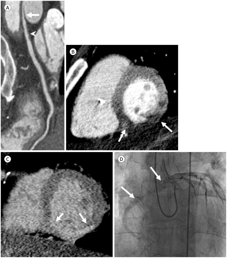
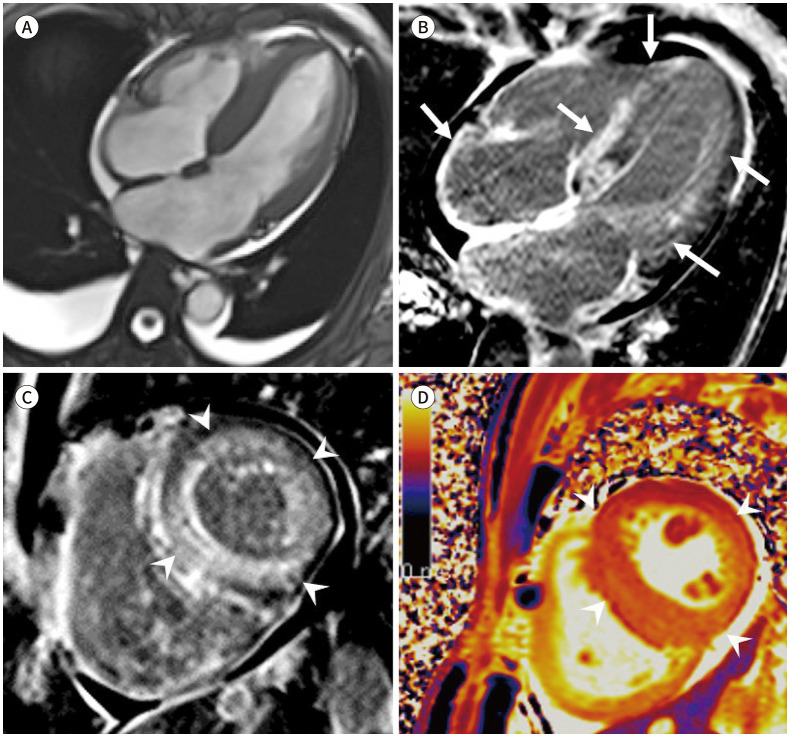
[CT and MR Imaging Findings of Structural Heart Diseases Associated with Sudden Cardiac Death]
- PMID: 36238400
- PMCID: PMC9432364
- DOI: 10.3348/jksr.2020.0161
[CT and MR Imaging Findings of Structural Heart Diseases Associated with Sudden Cardiac Death]
Abstract
Sudden cardiac death is an unexpected death originating from the heart that occurs within an hour of the onset of symptoms. The main cause of sudden cardiac death is arrhythmia; however, diagnosing underlying structural heart disease significantly contributes to predicting the long-term risk. Cardiovascular CT and MR provide important information for diagnosing and evaluating structural heart disease, enabling the prediction and preparation of the risk of sudden cardiac death. Therefore, we would like to focus on the various structural heart diseases that increase the risk of clinically-important sudden cardiac death and the importance of imaging findings.
급성 심장사는 증상이 시작된 후 한 시간 이내에 발생하는 심장 원인으로 인한 사망이다. 급성 심장사의 원인은 주로 부정맥이지만 동반할 수 있는 기저 심질환들을 사전에 진단하는 것은 장기적 위험을 예측하는 데 중요하다. 심장 CT와 심장 MR은 구조적 심질환을 진단하고 평가하는데 중요한 정보를 제공하여 급성 심장사의 위험을 예측하고 대비할 수 있게 한다. 따라서 임상적으로 중요한 급성 심장사의 위험을 증가시키는 다양한 원인과 영상 소견의 중요성에 대하여 중점적으로 살펴보고자 한다.
Copyrights © 2021 The Korean Society of Radiology.
Conflict of interest statement
Conflicts of Interest: The authors have no potential conflicts of interest to disclose.
Figures


References
-
- Zipes DP, Camm AJ, Borggrefe M, Buxton AE, Chaitman B, Fromer M, et al. ACC/AHA/ESC 2006 Guidelines for Management of Patients With Ventricular Arrhythmias and the Prevention of Sudden Cardiac Death: a report of the American College of Cardiology/American Heart Association Task Force and the European Society of Cardiology Committee for Practice Guidelines (writing committee to develop Guidelines for Management of Patients With Ventricular Arrhythmias and the Prevention of Sudden Cardiac Death): developed in collaboration with the European Heart Rhythm Association and the Heart Rhythm Society. Circulation. 2006;114:e385–e484. - PubMed
-
- Henneman MM, Schuijf JD, Jukema JW, Holman ER, Lamb HJ, De Roos A, et al. Assessment of global and regional left ventricular function and volumes with 64-slice MSCT: a comparison with 2D echocardiography. J Nucl Cardiol. 2006;13:480–487. - PubMed
-
- Salerno M, Beller GA. Noninvasive assessment of myocardial perfusion. Circ Cardiovasc Imaging. 2009;2:412–424. - PubMed
-
- Wu W, Budovec J, Foley WD. Prospective and retrospective ECG gating for thoracic CT angiography: a comparative study. AJR Am J Roentgenol. 2009;193:955–963. - PubMed
Publication types
LinkOut - more resources
Full Text Sources
