. 2022 May;83(3):597-607.
doi: 10.3348/jksr.2021.0186. Epub 2022 May 25.

[Mediastinal Teratoma: A Pictorial Essay]

[Article in Korean]

[Mediastinal Teratoma: A Pictorial Essay]

[Article in Korean]
Sang Gyun Kim et al. Taehan Yongsang Uihakhoe Chi. 2022 May.

Abstract

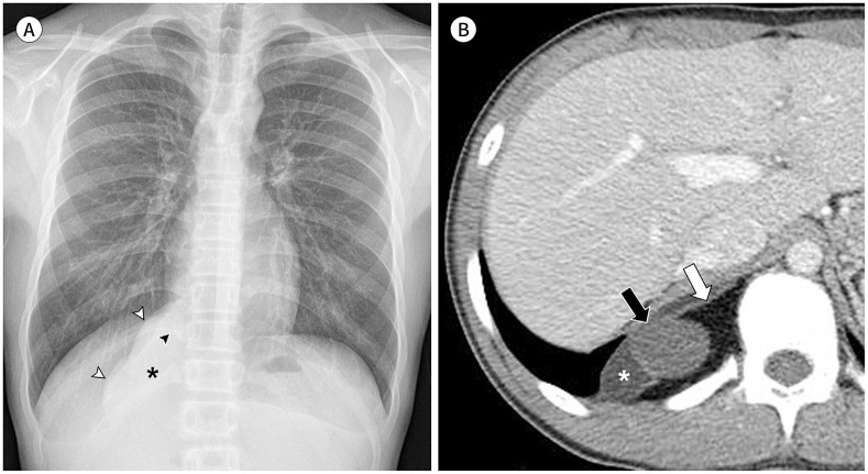
The mediastinum is the most prevalent site of extragonadal teratomas. Patients with mediastinal mature teratomas are usually young adults, and the condition does not show significant sexual differences. Mediastinal teratomas are mostly located in the anterior mediastinum. Patients are usually asymptomatic, although they can have several complications when the teratomas become large or rupture. Most mediastinal teratomas can be diagnosed using CT. Diagnosing ruptured or malignant teratomas is challenging because of their atypical clinical and radiological presentations. In this article, we describe various manifestations of mediastinal teratomas, with an emphasis on radiologic features.

종격동은 생식선 이외에서 발생하는 기형종의 가장 호발하는 부위로 알려져 있다. 종격동 성숙 기형종은 주로 젊은 성인에서 나타나고 남녀 간 발생 빈도의 큰 차이 없이 나타난다. 종격동 기형종은 대부분 전종격동에서 관찰되고, 일반적으로 증상을 보이지 않지만 종괴의 크기가 크거나 파열이 발생할 경우 여러 합병증이 나타날 수 있다. 종격동 기형종은 대부분 조직 검사 없이 전산화단층촬영(CT)만으로 진단될 수 있다. 하지만 파열된 기형종 혹은 악성 기형종의 경우 임상적 그리고 영상의학적으로 비전형적인 양상을 보여 진단이 어려울 수 있다. 본 종설에서는 종격동 기형종의 다양한 발현 양상을 영상 검사 소견을 중심으로 살펴보고자 한다.


Conflict of interest statement

Conflicts of Interest: Yun-Hyeon Kim has been a Editorial Board Member of the Journal of the Korean Society of Radiology since 2002; however, he was not involved in the peer reviewer selection, evaluation, or decision process of this article. Otherwise, no other potential conflicts of interest relevant to this article were reported.

Figures

Fig. 1
Fig. 1. A mediastinal mature teratoma in a 44-year-old-male.
A. Chest PA radiograph shows a mediastinal mass with curvilinear calcification (arrow), loss of anterior junction line (asterisk), and a hilum overlay sign (arrowhead). B. An axial contrast-enhanced CT scan shows a mass of approximately 10 cm with a calcified wall (open arrow) in the anterior mediastinum. The mass contains gross fat (asterisk), calcification (arrowhead), and areas of soft tissue (white arrow). C, D. In the thoracic MRI, soft tissue within the mass (arrows) has high signal intensity on a high b-value diffusion-weighted image (b = 800 mm2/s) (C) and low signal intensity on an apparent diffusion coefficient map (D), indicating diffusion restriction.
Fig. 2
Fig. 2. A cystic mediastinal mature teratoma in a 52-year-old male.
A. An axial contrast-enhanced CT scan shows an unilocular cystic mass in the right anterior mediastinum. B. An axial fat-suppressed T2-weighted image of an MRI reveals internal septation (arrowheads) and a soft tissue area (arrow) within the mass. C. Photomicrograph (hematoxylin and eosin stain, × 20) of the soft tissue area within a surgical resection specimen shows collagen material (asterisk), hair follicle (white arrow), adipose tissue (open arrow), and intestinal columnar epithelium (arrowheads).
Fig. 3
Fig. 3. A posterior mediastinal mature teratoma in a 12-year-old boy.
A. Chest PA shows a lobulating mass (asterisk) in the right paravertebral area. The mass displays negative a silhouette sign with abutting right atrium (black arrowhead) and right hemidiaphragm (white arrowheads), indicating posterior mediastinal location. B. An axial contrast-enhanced CT scan demonstrates a well-defined mass containing gross fat (white arrow), a cystic portion (black arrow), and an area of soft tissue (asterisk) in the right posterior mediastinum.
Fig. 4
Fig. 4. A ruptured mediastinal teratoma with empyema in a 45-year-old female presenting with chest pain and dyspnea.
A. A posteroanterior chest radiograph shows a large mass in the right middle and lower lung fields. The mass displays a silhouette sign with a right atrium (asterisk), raising the possibility of a mediastinal location. Pleural effusion (arrowheads) is also present in the right hemithorax. B, C. An axial contrast-enhanced CT scan shows a huge multiseptated cystic mass with areas of soft tissue (black arrows) in the right hemithorax and a right anterior mediastinum (asterisk). Loculated pleural effusion (arrowheads) is present, and focal wall dehiscence is observed between the cystic mass and pleural effusion (white arrow). Surgical resection was performed. Pus in the pleural cavity was visible during surgery, indicating empyema.
Fig. 5
Fig. 5. A ruptured mediastinal teratoma with pericarditis in a 19-year old male presenting with left chest pain and dyspnea.
A. An axial contrast-enhanced CT scan at the main pulmonary artery level shows a multilocular cystic and solid mass containing gross fat (arrow) in the anterior mediastinum. The mass demonstrates an indistinct margin, with increased opacity in the adjacent mediastinal fat. Also left pleural effusion is noted. B. A CT scan at the ventricle level reveals a small pericardial effusion with pericardial thickening and enhancement, indicating pericarditis. C. Axial CT at the brachiocephalic vein level. The mass encases the left brachiocephalic vein on the contrast-enhanced CT scan (arrowheads), raising the suspicion of vascular invasion. D. The fat-suppressed T2-weighted MR image reveals hyperintense wall thickening of the encasing left brachiocephalic vein, indicating acute inflammation (arrowheads).
Fig. 6
Fig. 6. Ruptured mediastinal teratoma with bronchial fistula in a 15-year-old girl who presented with hemoptysis.
A. An axial contrast-enhanced CT scan shows a lobulating cystic and solid mass containing gross fat (asterisk) in the left anterior mediastinum. Compressive atelectasis is observed in the adjacent left upper lobe (arrowheads). B, C. Lung window CT images demonstrate a fistula between the mass and the left upper lobe bronchus (arrow) (B) and extensive nodular ground-glass opacity in the scanned both lungs, indicating aspirated blood products (C).
Fig. 7
Fig. 7. A malignant mediastinal teratoma with malignant epithelial component in a 19-year-old male, with elevated human chorionic gonadotropin levels (54400 mIU/mL; 0–5) and a normal alpha fetoprotein level.
A. An axial contrast-enhanced CT scan shows a large soft tissue mass containing gross fat (asterisk) in the anterior mediastinum. Pulmonary metastases are also observed in both lungs (arrowheads). B. A CT scan at the upper abdomen level reveals renal metastasis in the right kidney (arrow). C. An axial contrast-enhanced T1-weighted image of the brain MRI shows brain metastases in the left occipital lobe (arrows).
Fig. 8
Fig. 8. A malignant mediastinal teratoma (mature teratoma with seminoma) in a 23-year-old male with elevated alpha fetoprotein levels (247.8 IU/mL; 0.74–7.29) and a normal human chorionic gonadotropin level.
A. An axial contrast-enhanced CT scan shows a cystic and solid mass in the anterior mediastinum. B. A coronal reformatting contrast-enhanced CT scan shows extracapsular tumoral growth at the superior portion of the mass (arrows).

References

    1. Jeung MY, Gasser B, Gangi A, Bogorin A, Charneau D, Wihlm JM, et al. Imaging of cystic masses of the mediastinum. Radiographics. 2002;22:S79–S93. - PubMed
    1. Hu X, Li D, Xia J, Wang P, Cai J. Mediastinal mixed germ cell tumor: a case report and literature review. Open Med (Wars) 2021;16:892–898. - PMC - PubMed
    1. Lewis BD, Hurt RD, Payne WS, Farrow GM, Knapp RH, Muhm JR. Benign teratomas of the mediastinum. J Thorac Cardiovasc Surg. 1983;86:727–731. - PubMed
    1. Shinagare AB, Jagannathan JP, Ramaiya NH, Hall MN, Van den Abbeele AD. Adult extragonadal germ cell tumors. AJR Am J Roentgenol. 2010;195:W274–W280. - PubMed
    1. Tian Z, Liu H, Li S, Chen Y, Ma D, Han Z, et al. Surgical treatment of benign mediastinal teratoma: summary of experience of 108 cases. J Cardiothorac Surg. 2020;15:36. - PMC - PubMed

Publication types