

CBCT-based assessment of root canal treatment using micro-CT reference images
- PMID: 36238704
- PMCID: PMC9530298
- DOI: 10.5624/isd.20220019
CBCT-based assessment of root canal treatment using micro-CT reference images
Abstract
Purpose: This study compared the root canal anatomy between cone-beam computed tomography (CBCT) and micro-computed tomography (micro-CT) images before and after biomechanical preparation and root canal filling.
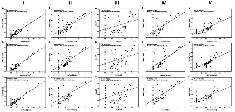
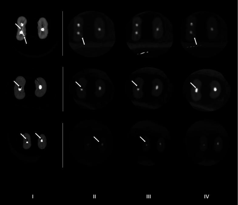
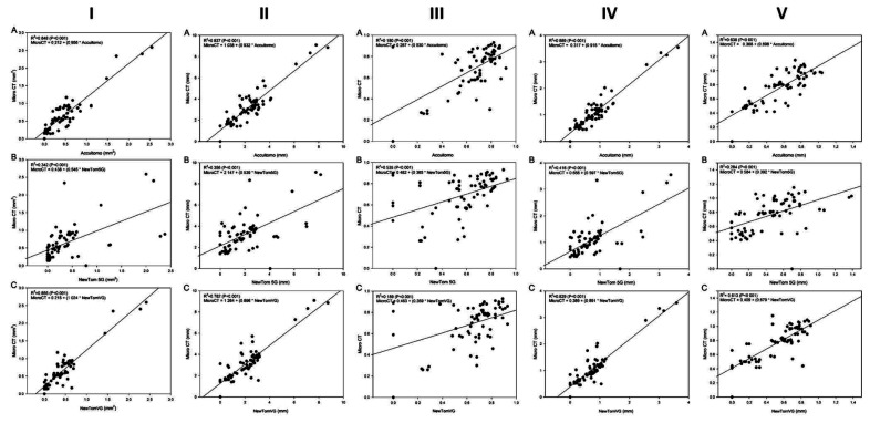
Materials and methods: Isthmus-containing mesial roots of mandibular molars (n=14) were scanned by micro-CT and 3 CBCT devices: 3D Accuitomo 170 (ACC), NewTom 5G (N5G) and NewTom VGi evo (NEVO). Two calibrated observers evaluated the images for 2-dimensional quantitative parameters, the presence of debris or root perforation, and filling quality in the root canal and isthmus. The kappa coefficient, analysis of variance, and the Tukey test were used for statistical analyses (α=5%).
Results: Substantial intra-observer agreement (κ=0.63) was found between micro-CT and ACC, N5G, and NEVO. Debris detection was difficult using ACC (42.9%), N5G (40.0%), and NEVO (40%), with no agreement between micro-CT and ACC, N5G, and NEVO (0.05<κ<0.12). After biomechanical preparation, 2.4%-4.8% of CBCT images showed root perforation that was absent on micro-CT. The 2D parameters showed satisfactory reproducibility between micro-CT and ACC, N5G, and NEVO (intraclass correlation coefficient: 0.60-0.73). Partially filled isthmuses were observed in 2.9% of the ACC images, 8.8% of the N5G and NEVO images, and 26.5% of the micro-CT images, with no agreement between micro-CT and ACC, and poor agreement between micro-CT and N5G and NEVO. Excellent agreement was found for area, perimeter, and the major and minor diameters, while the roundness measures were satisfactory.
Conclusion: CBCT images aided in isthmus detection and classification, but did not allow their classification after biomechanical preparation and root canal filling.
Keywords: Cone-Beam Computed Tomography; Endodontics; X-Ray Microtomography.
Copyright © 2022 by Korean Academy of Oral and Maxillofacial Radiology.
Conflict of interest statement
Conflicts of Interest: None
Figures


References
-
- Estrela C, Rabelo LE, de Souza JB, Alencar AH, Estrela CR, Sousa Neto MD, et al. Frequency of root canal isthmi in human permanent teeth determined by cone-beam computed tomography. J Endod. 2015;41:1535–1539. - PubMed
-
- Patel K, Mannocci F, Patel S. The assessment and management of external cervical resorption with periapical radiographs and cone-beam computed tomography: a clinical study. J Endod. 2016;42:1435–1440. - PubMed
-
- Beacham JT, Geist JR, Yu Q, Himel VT, Sabey KA. Accuracy of cone-beam computed tomographic image interpretation by endodontists and endodontic residents. J Endod. 2018;44:571–575. - PubMed
-
- Martins JN, Ordinola-Zapata R, Marques D, Francisco H, Caramês J. Differences in root canal system configuration in human permanent teeth within different age groups. Int Endod J. 2018;51:931–941. - PubMed
-
- Patel S, Brown J, Pimentel T, Kelly RD, Abella F, Durack C. Cone beam computed tomography in Endodontics - a review of the literature. Int Endod J. 2019;52:1138–1152. - PubMed
