Case Reports
. 2022 Sep 27:9:936949.
doi: 10.3389/fsurg.2022.936949. eCollection 2022.

Case report: Epidural blood patches are effective in treating intracranial hypotension due to a subarachnoid-pleural fistula

Affiliations
Case Reports

Case report: Epidural blood patches are effective in treating intracranial hypotension due to a subarachnoid-pleural fistula

Hua Huang et al. Front Surg. .

Abstract

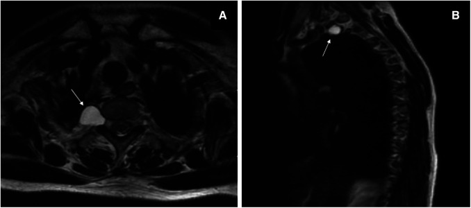
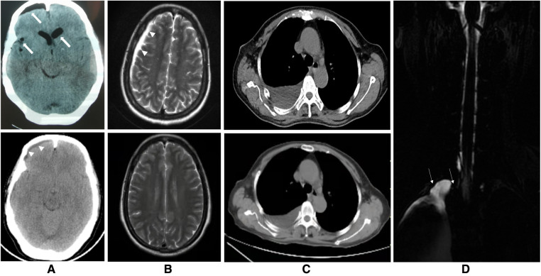
Background: Intracranial hypotension (IH) is usually associated with cerebrospinal fluid (CSF) leakage and/or CSF hypotension, and epidural blood patch (EBP) therapy has been proven to be effective for treating spontaneous IH and post-dural puncture headaches. Tarlov cysts (TCs) are common lesions of the sacral spine. They have rarely been reported in thoracic locations and are even less common in the posterior mediastinum, which can lead to their misdiagnosis as neurogenic tumors.

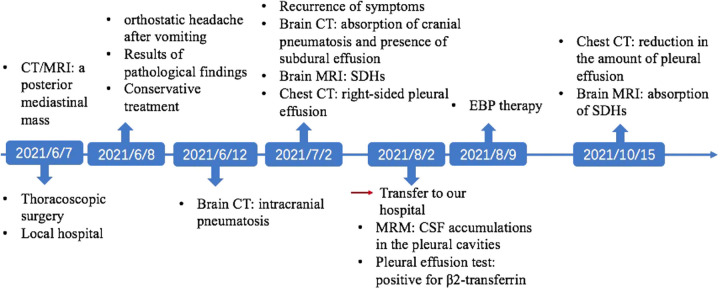
Case presentation: Here, we report the case of a 60-year-old woman who developed an orthostatic headache after the thoracoscopic resection of a TC in the posterior mediastinum that was presumed to be a schwannoma preoperatively. The patient was finally diagnosed with IH caused by a subarachnoid-pleural fistula (SPF) and was cured by targeted EBP treatment.

Conclusion: This is a case to show that a single targeted EBP treatment is effective for a patient with IH caused by an SPF after thoracoscopic resection of a TC. This case reminds us to be vigilant that a TC may be masquerading as a posterior mediastinal neurogenic tumor, and a detailed examination should be performed to identify it before deciding on a surgical procedure. In addition, postural headache after thoracoscopic spinal surgery should be alert to the possibility of IH induced by an SPF. Once it occurs, early treatment is necessary, and targeted EBP treatment can serve as a safe and effective alternative when conservative treatment fails.

Keywords: Tarlov cyst; epidural blood batch; intracranial hypotension; posterior mediastinal mass; subarachnoid-pleural fistula.


Conflict of interest statement

The authors declare that the research was conducted in the absence of any commercial or financial relationships that could be construed as a potential conflict of interest.

Figures

Figure 1
Figure 1
T2-weighted magnetic resonance imaging before the thoracoscopic surgery. Axial image (A) and sagittal image (B) showing a hyperintense oval lesion, with signal intensity similar to fluid, originating in the posterior mediastinum, protruding into the apical thorax at the level of the right T1–T2 intervertebral foramen (white arrows).
Figure 2
Figure 2
Images of the brain, chest and spine. (A) The upper figure of the axial computed tomography (CT) showing pneumocephalus in the cranial cavity of the lateral ventricle and right frontal, parietal and temporal and ventricular dilatation 5 days after the thoracoscopic surgery (thick arrows) and the lower figure showing resolution of these findings and presence of right frontal subdural effusion 25 days after the thoracoscopic surgery (arrowheads). (B) The upper figure of axial T2-weighted magnetic resonance imaging of the brain showing subdural hematomas in the left frontal region, right frontotemporal parietal occipital region before the epidural blood patch (EBP) therapy (arrowheads) and the lower figure showing complete absorption of these subdural hematomas 2 months after EBP therapy. (C) The upper figure of axial chest CT demonstrating a moderate amount of right-sided pleural effusion before the EBP therapy and the lower figure showing decrease of it 2 months after EBP therapy. (D) T2-weighted magnetic resonance myelography imaging of the spine showing subarachnoid-pleural fistula connection of the left pleural effusions and the spinal canal before EBP therapy (white arrow).
Figure 3
Figure 3
Timeline of the patient's clinical courses.

References

    1. Yoon SH, Chung YS, Yoon BW, Kim JE, Paek SH, Kim DG. Clinical experiences with spontaneous intracranial hypotension: a proposal of a diagnostic approach and treatment. Clin Neurol Neurosurg. (2011) 113(5):373–9. 10.1016/j.clineuro.2010.12.015 - DOI - PubMed
    1. Ferrante E, Trimboli M, Rubino F. Spontaneous intracranial hypotension: review and expert opinion. Acta Neurol Belg. (2020) 120(1):9–18. 10.1007/s13760-019-01166-8 - DOI - PubMed
    1. Loya JJ, Mindea SA, Yu H, Venkatasubramanian C, Chang SD, Burns TC. Intracranial hypotension producing reversible coma: a systematic review, including three new cases. J Neurosurg. (2012) 117(3):615–28. 10.3171/2012.4.JNS112030 - DOI - PubMed
    1. Cornips EM, Staals J, Stavast A, Rijkers K, van Oostenbrugge RJ. Fatal cerebral and cerebellar hemorrhagic infarction after thoracoscopic microdiscectomy: case report. J Neurosurg Spine. (2007) 6(3):276–9. 10.3171/spi.2007.6.3.276 - DOI - PubMed
    1. Shin HY. Recent update on epidural blood patch. Anesth Pain Med. (2022) 17(1):12–23. 10.17085/apm.21113 - DOI - PMC - PubMed

Publication types

LinkOut - more resources