Review
. 2023 Jan;29(1):e3457.
doi: 10.1002/psc.3457. Epub 2022 Nov 9.

Peptide-based covalent inhibitors of protein-protein interactions

Affiliations
Review

Peptide-based covalent inhibitors of protein-protein interactions

Felix M Paulussen et al. J Pept Sci. 2023 Jan.

Abstract

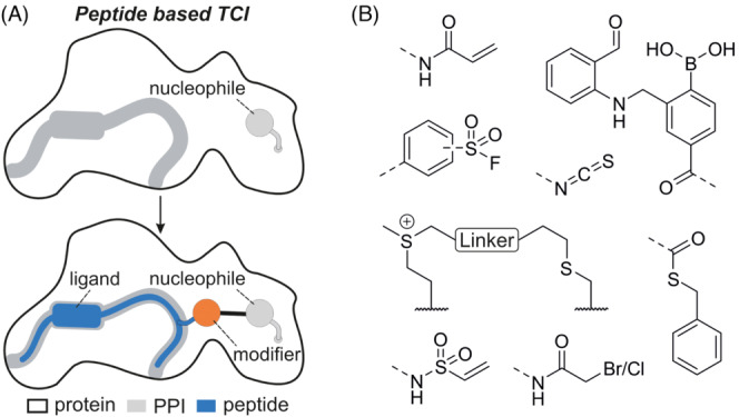
Protein-protein interactions (PPI) are involved in all cellular processes and many represent attractive therapeutic targets. However, the frequently rather flat and large interaction areas render the identification of small molecular PPI inhibitors very challenging. As an alternative, peptide interaction motifs derived from a PPI interface can serve as starting points for the development of inhibitors. However, certain proteins remain challenging targets when applying inhibitors with a competitive mode of action. For that reason, peptide-based ligands with an irreversible binding mode have gained attention in recent years. This review summarizes examples of covalent inhibitors that employ peptidic binders and have been tested in a biological context.

Keywords: bioconjugation; new modalities; peptidomimetics; proteomimetics; structure-based design.


Figures

FIGURE 1
FIGURE 1
(A) Structure‐based design of a peptide‐based targeted covalent inhibitor. Top: Ligand binding site (grey) in proximity to a nucleophile on the protein surface. Bottom: Introduction of a covalent modifier (orange) in the peptide ligand (blue) results in the desired covalent inhibitor. (B) Chemical structures of selected modifiers discussed in this review.
FIGURE 2
FIGURE 2
(A) Sequences and modification sites of PHYL‐derived peptides and covalent inhibitors of SIAH. Chemical structure of the tested acrylamide modifiers (x = 3 or 4) is provided. (B) Crystal structure of covalent inhibitor BI‐107F7 bound to SIAH C130 (pdb: 4i7d).
FIGURE 3
FIGURE 3
(A) Top: Crystal structure of NOXA BH3 (blue, pdb: 3mqp) and BIM BH3 (orange, pdb: 2vm6) bound to BFL‐1 (pdb: 3mqp). Bottom: Crystal structure of i, i + 7 stapled covalent inhibitor D‐NA‐NOXA SAHB bound to C55 of BFL‐1 (pdb: 5whh). (B) Sequences and modification sites of BFL‐1, BCL‐2A1 and MCL‐1 binding peptides. (C) Scheme of the different employed crosslinks. (D) Chemical structures of tested modifiers.
FIGURE 4
FIGURE 4
(A) Sequence of BIM BH3 and derived N‐capped peptides with introduced modifications (* α‐aminoisobutyric acid, Aib). (B) Chemical structure of N‐cap and chloroacetamide modifier in 138C5. (C) Crystal structure of 138C7 covalently bound to C55 of BFL‐1 (pdb: 2vm6). (D) Sequence of ephrin‐B PDZ binding domain/NCB1 and derived covalent inhibitor TAT‐CB‐6 with modification sites. TAT refers to a HIV TAT cell penetrating peptide (CPP).
FIGURE 5
FIGURE 5
(A) Sequence of MLL‐transactivation domain L and derived covalent binders with modification sites. (B) Chemical structures of modifiers (x = 1 or 3). (C) NMR structure (pdb: 2lxs) of KIX‐domain of CBP (white) and MLL‐transactivation domain (blue) with positions used for the generation of cysteine variants (beige). (D) NMR structure of GRB‐2 bound to a SOS‐1 derived peptide (pdb: 1gbq). (E) Sequence of SOS‐1‐derived peptide and derived monomeric and dimeric covalent inhibitors attached to a cell penetrating peptide (CPP).
FIGURE 6
FIGURE 6
(A) Sequence of wt SOS‐1 and derived inhibitors with modification sites (i: β‐isoleucine, e: β‐glutamic acid, a: β‐alanine, X: 4‐pentenoic acid, Z: 5‐hexenoic acid, z: N‐allylglycine). (B) Chemical structure of the hydrogen bond surrogate (HBS), selected β‐amino acids (bold), position of the modifier m and the HBS are shown. (C) Crystal structure of Ras (white, pdb: 1nvw) in complex with SOS derived peptide (wt SOS, blue, pdb: 1nvw, glycine was varied to cysteine for demonstration purposes). (D) Chemical structure of tested electrophiles (x = 1 or 2).
FIGURE 7
FIGURE 7
(A) Sequence of PDZ binding domain of ephrin‐B and derived sulfonium crosslinked peptide PD3 with introduced modifications. (B) AlphaFold, prediction of complex between ephrin‐B PDZ binding domain and PDZ. (C) Scheme of sulfonium bridged peptide. (D) Chemical structure of linkers used for crosslinking of ephrin‐B. Only the linkers on the left side were employed in the crosslinking of BIM BH3. (E) Sequence of BIM BH3 and derived sulfonium crosslinked peptide B4‐MCI with location of introduced modifications (*α‐aminoisobutyric acid, Aib).
FIGURE 8
FIGURE 8
(A) Crystal structure of gp41 helix bundle (pdb: 1szt) with C‐terminal heptad repeat (CHR, blue) and N‐terminal heptad repeat (NHR, beige). (B) Sequence of C34 and derived inhibitors with modifications. SC35E(SBn) 5 H 9 and SC22E(SBn) 5 H 9 have been altered introducing additional salt bridges to stabilize helicity and solubility. The bold histidine residue has been introduced to increase NHR K574 reactivity. C corresponds to a C‐terminally attached cholesterol. (C) Chemical structure of the employed modifiers and their reactivity.
FIGURE 9
FIGURE 9
(A) Crystal structure of Mdm4 with p53 transactivation domain (pdb: 3dac). (B) Crystal structure of Mdm2 with SAHp53–8 (pdb: 3v3b). (C) Sequence of p53 (14‐29) and derived inhibitors with modifications. (D) Scheme of i, i + 7 stapled peptide. (E) Chemical structure of tested modifiers (x = 1, 2, 3).
FIGURE 10
FIGURE 10
(A) Crystal structure of BIR3 domain of XIAP with SMAC derived peptide AVPF (pdb: 2opz). (B) Sequence of SMAC wt and derived inhibitors with modification sites. (C) Chemical structure of a selection of tested modifiers (x = 1, 2, 3). (D) Chemical structure of N‐terminal modifications.
FIGURE 11
FIGURE 11
(A) Crystal structure of MCL‐1 (white, pdb: 6vbx) in complex with BIM BH3 peptide (orange, pdb: 2 nl9) or covalent inhibitor 15 (beige, pdb: 6vbx). (B) Sequence of BIM BH3 and derived MCL‐1 inhibitors with modification sites. Additional salt bridges were introduced (bold) to increase helicity. (C) Chemical structure of selected modifiers (x = 1, 2, 3).
FIGURE 12
FIGURE 12
(A) Sequences of reversible covalent inhibitors. (B) Scheme of cyclized W7‐derived inhibitors. (C) Chemical structures of selected reversible modifiers in their bound and unbound state. (D) Scheme of cyclized binders of SrtA and spike RBD including chemical structures of crosslink and modifier APBA‐3.
FIGURE 13
FIGURE 13
(A) Sequence of FtsB and different FtsB derived peptides (B: Norleucine). (B) Scheme of cyclized FtsB‐derived peptides including chemical structure of hydrocarbon crosslink. (C) MD simulation of 24f bound to FtsQ. (D) Chemical structures of tested modifiers.

References

    1. Amaral LAN. A truer measure of our ignorance. Proc Natl Acad Sci USA. 2008;105(19):6795‐6796. doi:10.1073/pnas.0802459105 - DOI - PMC - PubMed
    1. Rolland T, Taşan M, Charloteaux B, et al. A proteome‐scale map of the human interactome network. Cell. 2014;159(5):1212‐1226. doi:10.1016/j.cell.2014.10.050 - DOI - PMC - PubMed
    1. De Las Rivas J, Fontanillo C. Protein–protein interactions essentials: key concepts to building and analyzing Interactome networks. PLoS Comput Biol. 2010;6(6):e1000807. doi:10.1371/journal.pcbi.1000807 - DOI - PMC - PubMed
    1. Hopkins AL, Groom CR. The druggable genome. Nat Rev Drug Discov. 2002;1(9):727‐730. doi:10.1038/nrd892 - DOI - PubMed
    1. Milroy L‐G, Grossmann TN, Hennig S, Brunsveld L, Ottmann C. Modulators of protein–protein interactions. Chem Rev. 2014;114(9):4695‐4748. doi:10.1021/cr400698c - DOI - PubMed

MeSH terms

LinkOut - more resources