. 2022 Nov;62(11):2334-2348.
doi: 10.1111/trf.17137. Epub 2022 Oct 14.

Anti-CD38 monoclonal antibody interference with blood compatibility testing: Differentiating isatuximab and daratumumab via functional epitope mapping

Affiliations

Anti-CD38 monoclonal antibody interference with blood compatibility testing: Differentiating isatuximab and daratumumab via functional epitope mapping

Btissam Chami et al. Transfusion. 2022 Nov.

Abstract

Background: There are two FDA-approved anti-CD38 monoclonal antibodies for treatment of multiple myeloma: isatuximab and daratumumab. Owing to expression of CD38 on reagent red blood cells (RBCs), these antibodies interfere with indirect antiglobulin tests (IATs). We sought to understand differences in such interference by performing binding experiments.

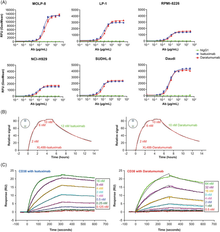
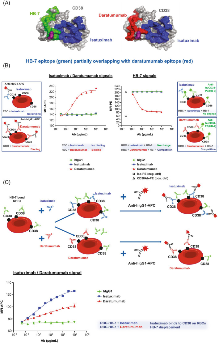
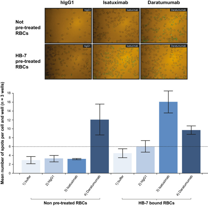
Study design and methods: In vitro experiments to compare the binding to RBCs of isatuximab and daratumumab alone or in the presence of a mouse anti-human CD38 antibody (HB-7 or AT13/5) or a nicotinamide adenine dinucleotide-analog CD38 inhibitor were performed and quantified by flow cytometry, imaging, mass spectrometry, surface plasmon resonance, and LigandTracer technologies. Serologic testing was performed on plasma samples spiked with isatuximab or daratumumab.

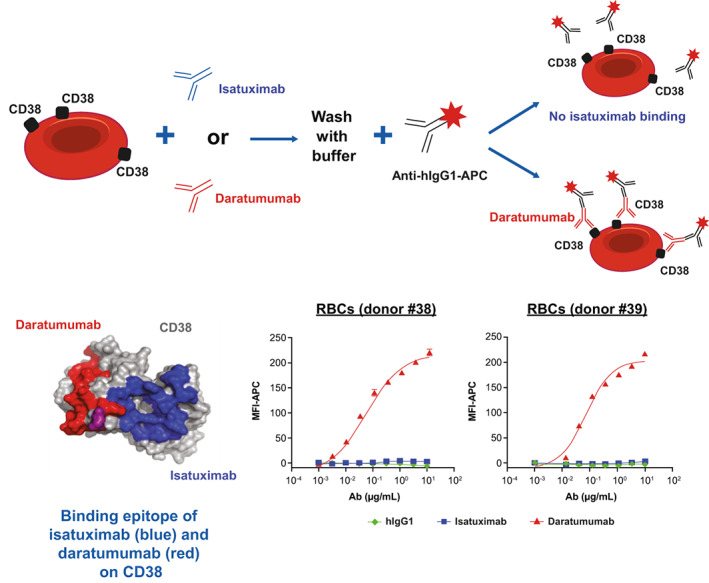
Results: CD38 expressed on RBCs can be directly bound by daratumumab, whereas isatuximab requires a co-factor, such as HB-7, AT13/5, or a CD38 inhibitor, suggesting that the isatuximab epitope on RBCs is masked in vitro. Daratumumab samples more frequently showed interference and had stronger reactions than isatuximab samples. Dithiothreitol treatment was equally effective in mitigating the interference caused by either drug.

Discussion: Both isatuximab and daratumumab interfere with IATs but at different magnitudes, reflecting distinct binding to CD38 on RBCs. From the binding studies, we conclude that the isatuximab epitope on RBCs is masked in vitro and binding requires a certain CD38 conformation or co-factor. This circumstance may explain why interference is seen only in a subset of patients receiving isatuximab when compared with interference seen in most patients on daratumumab therapy.


Conflict of interest statement

Btissam Chami: No relevant financial relationships to disclose. Makoto Okuda: No relevant financial relationships to disclose. Morvarid Moayeri: Institution support—Sanofi. France Pirenne: No relevant financial relationships to disclose. Yoko Hidaka: No relevant financial relationships to disclose. Ashok Nambiar: Institution support—Sanofi. Zhili Song: Employed by Sanofi; may hold stock and/or stock options in the company. Olivier Bedel: Employed by Sanofi at the time of study. Bailin Zhang: Employed by Sanofi; may hold stock and/or stock options in the company. Joern Hopke: Employed by Sanofi; may hold stock and/or stock options in the company. Gejing Deng: Employed by Sanofi; may hold stock and/or stock options in the company. Chen Zhu: Employed by Sanofi; may hold stock and/or stock options in the company. Sandrine Macé: Employed by Sanofi; may hold stock and/or stock options in the company. Marielle Chiron: Employed by Sanofi; may hold stock and/or stock options in the company. Francisco Adrian: Employed by Sanofi at the time of study. Taro Fukao: Employed by Sanofi; may hold stock and/or stock options in the company. Frank Basile: Employed by Sanofi at the time of study. Thomas Martin: Research funding—Sanofi, Seattle Genetics, Amgen, Janssen, GSK.

Figures

FIGURE 1
FIGURE 1
Isatuximab and daratumumab binding to CD38 expressed on cancer cell lines and rhCD38. (A) Detection of surface‐bound anti‐CD38 antibodies on MM cells (MOLP‐8, LP‐1, RPMI‐8226, NCI‐H929) or B‐cell lymphoma (SUDHL‐8, Daudi) by flow‐cytometric analysis. Cells (0.2 E6) were treated with hIgG1 (green), isatuximab (blue), or daratumumab (red) at concentrations ranging from 0–30 μg/ml. Surface‐bound antibody was detected with FITC‐Goat F(ab’)2 anti‐hIgG. (B) Determination of binding kinetics and binding affinity using LT. LT experiments were conducted with cells attached to the detection spots (small circles with “+” for JJN3‐CD38+ cells and “‐” for JJN3 parental cells) on a petri dish (schematically shown as a large circle on the top left of each time traces). Association and dissociation time traces (black, background and reference subtracted) are fitted with a 1:1 kinetic model (red). Concentrations of anti‐CD38 monoclonal antibody labeled with XL488 (XL488‐Isatuximab or XL488‐Daratumumab in red) added for association and unlabeled antibody (isatuximab or daratumumab in green) added for dissociation are shown in the figures. (C) Determination of binding kinetics and binding affinity using SPR. SPR experiments were conducted with isatuximab and daratumumab captured by anti‐Fc antibody immobilized on CM5 sensor chip. rhCD38 at varied concentrations (0.125–16.0 nM for isatuximab and 0.5–64 nM for daratumumab) was injected over the sensor surface. Association and dissociation time traces (colored, background, and reference subtracted) are fitted with a 1:1 kinetic binding model (black). K D values are calculated based on k a and k d from the fits of the binding kinetics. Ab, antibody; hIgG1, human immunoglobulin G1; ka, association rate constant; kd, dissociation rate constant; K D, equilibrium dissociation constant; LT, LigandTracer; rhCD38, recombinant human CD38; SPR, surface plasmon resonance; RFU, relative fluorescence unit; RU, response unit [Color figure can be viewed at wileyonlinelibrary.com]
FIGURE 2
FIGURE 2
In vitro binding of isatuximab and daratumumab to RBCs. RBCs from healthy donors were treated with hIgG1 (green), isatuximab (blue), or daratumumab (red) at concentrations ranging from 0 to 10 μg/ml. Surface‐bound anti‐CD38 antibody was detected with goat anti‐hIgG‐APC by flow cytometry. Ab, antibody; APC, allophycocyanin; hIgG1, human immunoglobulin G1; MFI, median fluorescent intensity; RBC, red blood cell [Color figure can be viewed at wileyonlinelibrary.com]
FIGURE 3
FIGURE 3
Daratumumab binding to CD38 on RBCs can be displaced by HB‐7 and pre‐treatment with HB‐7 induces binding of isatuximab to RBCs. (A) Representation of CD38 antigenic determinants of isatuximab (blue), daratumumab (red), and HB‐7 (green). E233 (purple) is a residue shared by all three epitopes. Protein Data Bank 4CMH (i.e., crystal structure of CD38 with CD38‐targeting antibody isatuximab) was used to generate CD38 surface. (B) Detection of APC signals from hIgG1 (green), isatuximab (blue), or daratumumab (red) binding to CD38 expressed on RBCs in vitro (left) or the ability of HB‐7 (PE signal) to bind CD38 expressed on RBCs in vitro in the presence of hIgG1 (green), isatuximab (blue), or daratumumab (red) (right) by flow cytometry. (C) RBCs were pre‐treated with mouse HB‐7 Ab before incubation with hIgG1 (green), isatuximab (blue) or daratumumab (red) at 0–100 μg/ml. Binding of isatuximab and daratumumab to CD38/HB‐7 complex on RBCs was detected with APC conjugated goat anti‐hIgG by flow cytometry. APC, allophycocyanin; ctrl, control; hIgG1, human immunoglobulin G1; MFI, median fluorescent intensity; PE, phycoerythrin; RBC, red blood cell [Color figure can be viewed at wileyonlinelibrary.com]
FIGURE 4
FIGURE 4
Confocal imaging analysis confirms HB‐7‐induced isatuximab binding to HB‐7‐bound RBCs. Binding of isatuximab and daratumumab (10 μg/ml) to RBCs in the absence or presence of HB‐7 (1 μg/ml) was evaluated by image‐based analysis. Green dots showing binding signals (AF488) on cell surface were visualized by microscope (60xW) (top panel of images) and number of positive spots were quantified (bottom graph). hIgG1, human immunoglobulin G1; RBC, red blood cell [Color figure can be viewed at wileyonlinelibrary.com]
FIGURE 5
FIGURE 5
In vitro assay, mouse anti‐human CD38 antibodies (HB‐7 or AT13/5) and CD38 inhibitor (ara‐F‐NAD) can induce isatuximab binding to CD38 on RBCs and increase isatuximab binding affinity for rhCD38. (A) Induced isatuximab binding to RBCs. Pre‐treatment of RBCs with mIgG, HB‐7, AT13/5, or CD38 inhibitor at indicated concentrations and detection of hIgG1 (green), isatuximab (blue), or daratumumab (red) by flow cytometry. Top: flow cytometry histograms; Bottom: dose curves. (B–C) Isatuximab binding kinetics and binding affinity to rhCD38 in the presence of HB‐7 and CD38 inhibitor were measured by SPR. Ab, antibody; APC, allophycocyanin; ara‐F‐NAD, ara‐F‐nicotinamide adenine dinucleotide; Comp, compensated; hIgG1, human immunoglobulin G1; K D, equilibrium dissociation constant; MFI, mean fluorescent intensity; mIgG, mouse immunoglobulin G; RBC, red blood cell; rhCD38, recombinant human CD38; SPR, surface plasmon resonance; RU, response unit; s, seconds [Color figure can be viewed at wileyonlinelibrary.com]

References

    1. Kazandjian D. Multiple myeloma epidemiology and survival: a unique malignancy. Semin Oncol. 2016;43(6):676–81. - PMC - PubMed
    1. Lin MH, Liu FY, Wang HM, Cho HC, Lo SC. Interference of daratumumab with pretransfusion testing, mimicking a high‐titer, low avidity like antibody. Asian J Transfus Sci. 2017;11(2):209–11. - PMC - PubMed
    1. Jiang H, Acharya C, An G, Zhong M, Feng X, Wang L, et al. SAR650984 directly induces multiple myeloma cell death via lysosomal‐associated and apoptotic pathways, which is further enhanced by pomalidomide. Leukemia. 2016;30(2):399–408. - PubMed
    1. Martin TG, Corzo K, Chiron M, Velde HV, Abbadessa G, Campana F, et al. Therapeutic opportunities with pharmacological inhibition of CD38 with isatuximab. Cell. 2019;8(12):1522. - PMC - PubMed
    1. Sarclisa (isatuximab‐irfc) injection for intravenous use . Prescribing information. sanofi‐aventis U.S. LLC. Available from: https://products.sanofi.us/Sarclisa/sarclisa.pdf. Accessed July 21, 2022.

Publication types