

Cerebrospinal fluid biomarker panel of synaptic dysfunction in Alzheimer's disease and other neurodegenerative disorders
- PMID: 36239248
- PMCID: PMC10102247
- DOI: 10.1002/alz.12809
Cerebrospinal fluid biomarker panel of synaptic dysfunction in Alzheimer's disease and other neurodegenerative disorders
Abstract
Introduction: Synaptic degeneration is a key part of the pathophysiology of neurodegenerative diseases, and biomarkers reflecting the pathological alterations are greatly needed.
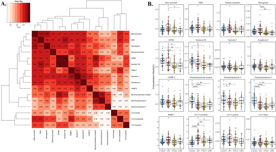
Method: Seventeen synaptic proteins were quantified in a pathology-confirmed cerebrospinal fluid cohort of patients with Alzheimer's disease (AD; n = 63), frontotemporal lobar degeneration (FTLD; n = 53), and Lewy body spectrum of disorders (LBD; n = 21), as well as healthy controls (HC; n = 48).
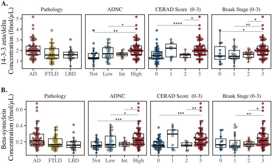
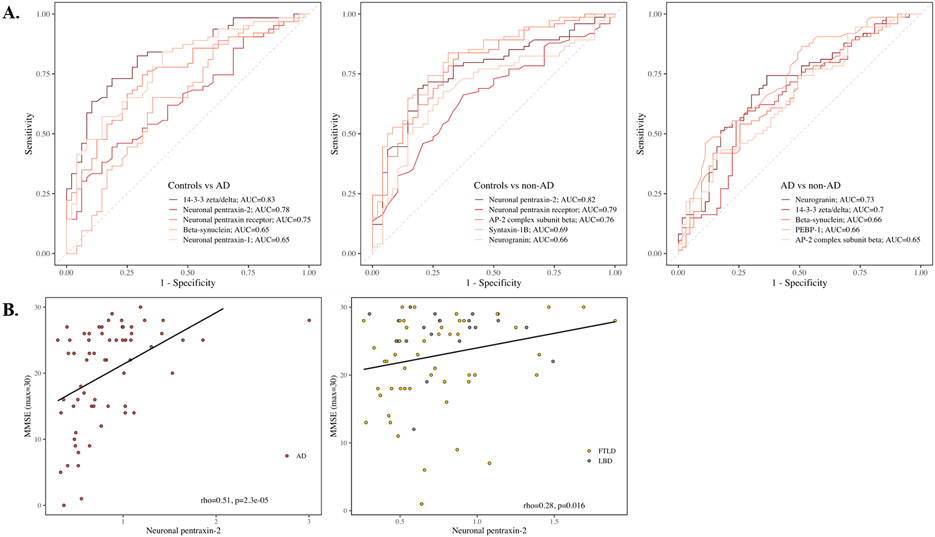
Results: Comparisons revealed four distinct patterns: markers decreased across all neurodegenerative conditions compared to HC (the neuronal pentraxins), markers increased across all neurodegenerative conditions (14-3-3 zeta/delta), markers selectively increased in AD compared to other neurodegenerative conditions (neurogranin and beta-synuclein), and markers selectively decreased in LBD and FTLD compared to HC and AD (AP2B1 and syntaxin-1B).
Discussion: Several of the synaptic proteins may serve as biomarkers for synaptic dysfunction in AD, LBD, and FTLD. Additionally, differential patterns of synaptic protein alterations seem to be present across neurodegenerative diseases.
Highlights: A panel of synaptic proteins were quantified in the cerebrospinal fluid using mass spectrometry. We compared Alzheimer's disease, frontotemporal degeneration, and Lewy body spectrum of disorders. Pathology was confirmed by autopsy or familial mutations. We discovered synaptic biomarkers for synaptic degeneration and cognitive decline. We found differential patterns of synaptic proteins across neurodegenerative diseases.
Keywords: Alzheimer's disease; Lewy body spectrum of disorders; biomarkers; frontotemporal lobar degeneration; mass spectrometry; synaptic pathology.
© 2022 The Authors. Alzheimer's & Dementia published by Wiley Periodicals LLC on behalf of Alzheimer's Association.
Figures
References
-
- Terry RD, et al. , Physical basis of cognitive alterations in Alzheimer's disease: synapse loss is the major correlate of cognitive impairment. Annals of Neurology: Official Journal of the American Neurological Association and the Child Neurology Society, 1991. 30(4): p. 572–580. - PubMed
-
- Davidsson P, Puchades M, and Blennow K, Identification of synaptic vesicle, pre- and postsynaptic proteins in human cerebrospinal fluid using liquid-phase isoelectric focusing. Electrophoresis, 1999. 20(3): p. 431–437. - PubMed
Publication types
MeSH terms
Substances
Grants and funding
LinkOut - more resources
Full Text Sources
Medical
