. 2022 Nov 1;149(21):dev200909.
doi: 10.1242/dev.200909. Epub 2022 Oct 31.

CXCL12 defines lung endothelial heterogeneity and promotes distal vascular growth

Affiliations

CXCL12 defines lung endothelial heterogeneity and promotes distal vascular growth

Prashant Chandrasekaran et al. Development. .

Abstract

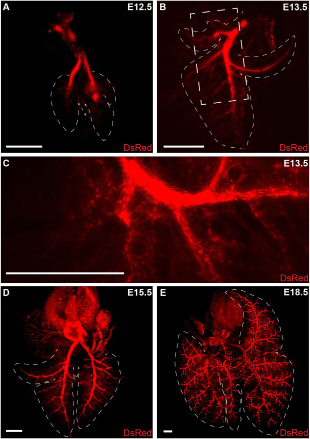
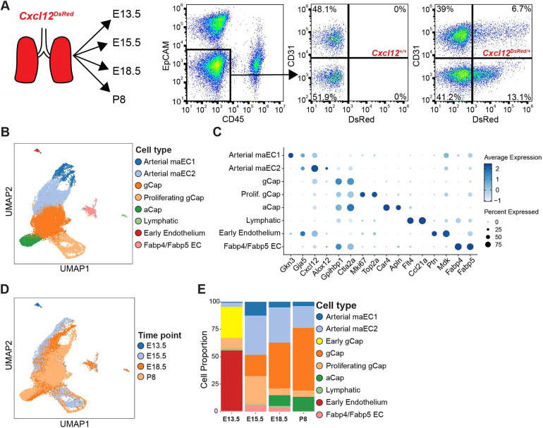
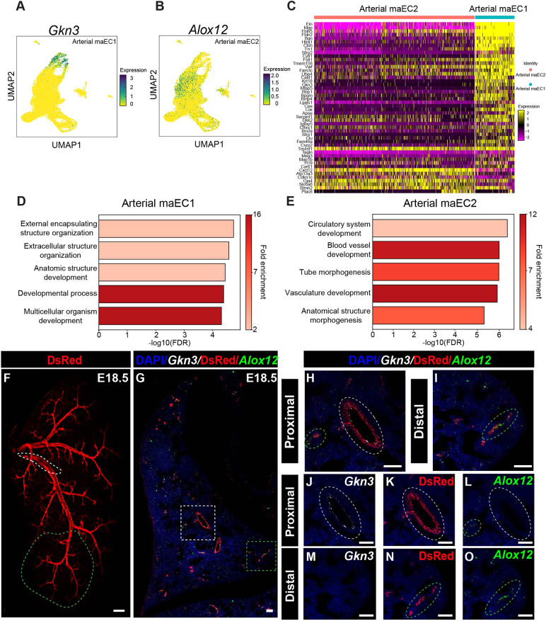
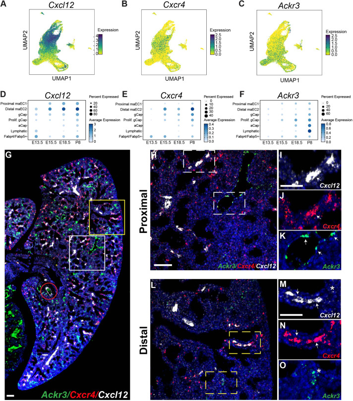
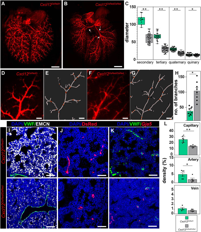
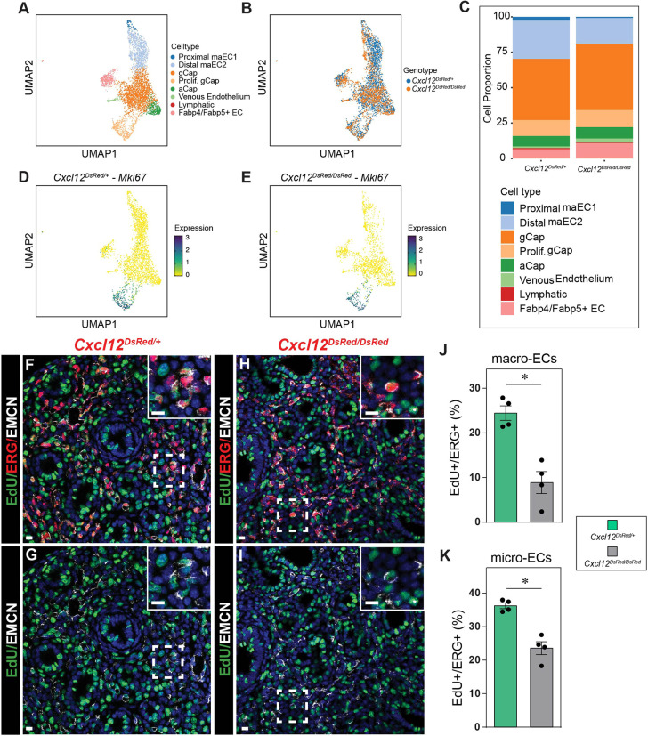
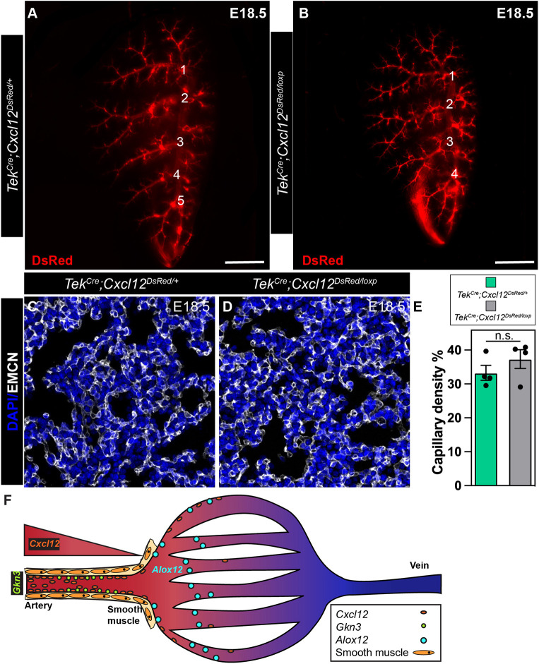
There is a growing amount of data uncovering the cellular diversity of the pulmonary circulation and mechanisms governing vascular repair after injury. However, the molecular and cellular mechanisms contributing to the morphogenesis and growth of the pulmonary vasculature during embryonic development are less clear. Importantly, deficits in vascular development lead to significant pediatric lung diseases, indicating a need to uncover fetal programs promoting vascular growth. To address this, we used a transgenic mouse reporter for expression of Cxcl12, an arterial endothelial hallmark gene, and performed single-cell RNA sequencing on isolated Cxcl12-DsRed+ endothelium to assess cellular heterogeneity within pulmonary endothelium. Combining cell annotation with gene ontology and histological analysis allowed us to segregate the developing artery endothelium into functionally and spatially distinct subpopulations. Expression of Cxcl12 is highest in the distal arterial endothelial subpopulation, a compartment enriched in genes for vascular development. Accordingly, disruption of CXCL12 signaling led to, not only abnormal branching, but also distal vascular hypoplasia. These data provide evidence for arterial endothelial functional heterogeneity and reveal conserved signaling mechanisms essential for pulmonary vascular development.

Keywords: Endothelium; Lung; Mouse; Single-cell RNA sequencing.


Conflict of interest statement

Competing interests L.R.Y. declares grants paid to her institution from the National Institutes of Health and the Orphan Disease Center at the University of Pennsylvania; royalties from UpToDate; consultancy fees and fees for participation on the Steering Committee of the InPedILD trial from Boehringer Ingelheim; and consultancy fees from Roche and Sanofi.

Figures

Fig. 1.
Fig. 1.
Morphogenesis of the developing pulmonary arterial tree in Cxcl12DsRed/+ reporter embryos. (A) At E12.5, two intrapulmonary arteries are visible. (B) At E13.5, secondary branches extend out from the primary branches. (C) Higher magnification of secondary branching (boxed area in B). (D) At E15.5, increased secondary branching can be observed along with tertiary and quaternary branches. (E) At E18.5, robust and complex branching is apparent. Dashed lines delineate outlining of the lung lobes. Scale bars: 500 µm.
Fig. 2.
Fig. 2.
Sequencing of Cxcl12+ cells uncovers heterogeneity and previously undefined arterial EC subpopulations. (A) FACS strategy and plots for Cxcl12-DsRed+ cells. (B) UMAP embedding of Cxcl12-DsRed+ cells (n=26,652) colored by cell type. (C) Dot plot of marker genes for each cell type where dot size indicates the proportion of cells within a cluster expressing a gene, and color intensity indicates the relative expression level. (D) UMAP embedding colored by time point. (E) Cellular composition changes in Cxcl12+ ECs over time.
Fig. 3.
Fig. 3.
Identification of proximal and distal arterial ECs by RNA FISH and predicted function analysis. (A) UMAP embedding of cells colored by Gkn3 expression. (B) UMAP embedding of cells colored by Alox12 expression. (C) Heat map representing expression of hallmark genes in arterial macro endothelial cell types 1 and 2 (arterial maEC1 and maEC2). (D) GO analysis of the maEC1 cluster. (E) GO analysis of the maEC2 cluster. (F) Whole-mount image showing Cxcl12-DsRed arterial endothelium. Dashed white line marks the proximal artery branch and the dashed green line marks the arterial tree from the distal tip of the secondary branch to the quinary branches. (G) RNA FISH for Gkn3 and Alox12 and IHC for DsRed protein at E18.5 proximal (white box) and distal (red box) regions. (H) Higher magnification of RNA FISH for Gkn3 and Alox12 and IHC for DsRed protein in the proximal region. (I) Higher magnification of RNA FISH for Gkn3 and Alox12 and IHC for DsRed protein in the distal region. (J-L) Separate channels for proximal region RNA FISH for Gkn3 (J), IHC staining for DsRed (K) and Alox12 (L). (M-O) Separate channels for proximal region RNA FISH for Gkn3 (M), IHC staining for DsRed (N) and Alox12 (O). Scale bars: 50 mm (F); 50 μm (G-O).
Fig. 4.
Fig. 4.
Characterization of Cxcl12 expression and its signaling axis. (A-C) UMAP embedding of cells colored by Cxcl12 (A), Cxcr4 (B) and Ackr3 (C) expression. (D-F) Dot plots of Cxcl12 (D) Cxcr4 (E) and Ackr3 (F) expression in different EC compartments at E13.5, E15.5, E18.5 and P8. (G) RNA FISH for spatial expression of Ackr3, Cxcr4 and Cxcl12 in ECs and VSMCs at E18.5. The red circle denotes a proximal artery. (H) Higher magnification of RNA FISH for Ackr3, Cxcr4 and Cxcl12 in proximal lung at E18.5 (white boxed area in G). (I-K) Higher magnification of the boxed areas in H. (L) Higher magnification of RNA FISH for Ackr3, Cxcr4 and Cxcl12 in distal lung at E18.5 (yellow boxed area in G). (M-O) Higher magnification of boxed areas in L. Arrows indicate expression in macrovessels. Asterisks indicate expression in capillaries. Scale bars: 100 µm (G); 50 µm (H-O).
Fig. 5.
Fig. 5.
Loss of Cxcl12 leads to branching defects and pulmonary vascular hypoplasia. (A,B) Whole-mount imaging of arterial endothelium in Cxcl12DsRed/+ (A) and Cxcl12DsRed/DsRed (B) at E18.5. Cxcl12DsRed/DsRed lungs show defective proximal branching (white arrows). (C) Arterial diameter distribution of control Cxcl12DsRed versus null Cxcl12DsRed/DsRed arteries (n=4, filaments measured ≥10, **P<0.005, *P<0.05). (D-H) Branching analysis using Imaris Filament. (D,E) Representative image of Cxcl12DsRed/+ artery (D) and Filament tracing using IMARIS 9.2 (E). (F,G) Representative image of Cxcl12DsRed/DsRed artery (F) and Filament tracing (G). (H) Number of branches from the quaternary branch in the periphery (n=4, images analyzed ≥8, *P<0.05). (I-L). Vascular density measurements in control Cxcl12DsRed (top) versus null Cxcl12DsRed/DsRed (bottom) lung tissue. (I) Capillary density IHC staining of tissue sections for VWF (macrovessel) and EMCN (capillary and vein). (J) Arterial density IHC staining of tissue sections for DsRed (artery). (K) Venous density IHC and RNA FISH staining for Gja5 (artery) and VWF (macrovessel) expression. (L) Volume density calculations of capillaries, arteries and veins (n≥4, **P<0.005, *P<0.05). Scale bars: 1 mm (A,B); 100 µm (D-G); 50 µm (I-K).
Fig. 6.
Fig. 6.
Decreased proliferation in control Cxcl12DsRed versus null Cxcl12DsRed/DsRed ECs. (A) UMAP embedding of cells colored by endothelial cell populations at E18.5. (B) UMAP embedding of cells colored by Cxcl12DsRed/+ and Cxcl12DsRed/DsRed ECs at E18.5. (C) Proportion of different cell types in Cxcl12DsRed/+ and Cxcl12DsRed/DsRed at E18.5. (D,E) UMAP embedding of cells colored by Mki67 expression in Cxcl12DsRed/+ (D) and Cxcl12DsRed/DsRed (E) ECs at E18.5. (F-K) Assessment of proliferation measured by EdU incorporation in Cxcl12DsRed/+ and Cxcl12DsRed/DsRed macro- and micro-ECs. (F-I) IHC for EdU, ERG and EMCN to mark proliferating macro- and micro-ECs in in Cxcl12DsRed/+ (F,G) and Cxcl12DsRed/DsRed (H,I) lung sections. Insets show higher magnification of the boxed areas. (J,K) Quantification of the percentage of EdU+/ERG+ macro- (J) and micro- (K) ECs in Cxcl12DsRed/+ and Cxcl12DsRed/DsRed lungs (n=4). *P<0.005. Scale bar: 10 µm (F-I, main panels); 10 µm (F-I, insets).
Fig. 7.
Fig. 7.
EC-specific loss of CXCL12 leads to branching defects. (A,B) Whole-mount imaging of the left lobe of TekCre;Cxcl12DsRed/+ (A) and TekCre;Cxcl12DsRed/loxp/+ (B) embryos (numbers signify branching points). (C,D) IHC staining for EMCN to delineate capillaries in TekCre;Cxcl12DsRed/+ (C) and TekCre;Cxcl12DsRed/loxp/+ (D) embryonic lungs. (E) Quantification of capillary density (n=4). n.s., not significant (P=0.485). (F) Model of CXCL12 and arterial heterogeneity. A gradient of Cxcl12 expression exists in distal pulmonary vasculature that delineates the arterial endothelium into proximal Gkn+ and distal Alox12+ subpopulation with distinct functions. Scale bars: 1 mm (A,B); 50 µm (C,D).

References

    1. Ahlmann-Eltze, C. and Huber, W. (2021). glmGamPoi: fitting Gamma-Poisson generalized linear models on single cell count data. Bioinformatics 36, 5701-5702. 10.1093/bioinformatics/btaa1009 - DOI - PMC - PubMed
    1. Ashburner, M., Ball, C. A., Blake, J. A., Botstein, D., Butler, H., Cherry, J. M., Davis, A. P., Dolinski, K., Dwight, S. S., Eppig, J. T.et al. (2000). Gene ontology: tool for the unification of biology. The Gene Ontology Consortium. Nat. Genet. 25, 25-29. 10.1038/75556 - DOI - PMC - PubMed
    1. Asosingh, K., Comhair, S., Mavrakis, L., Xu, W., Horton, D., Taylor, I., Tkachenko, S., Hu, B. and Erzurum, S. (2021). Single-cell transcriptomic profile of human pulmonary artery endothelial cells in health and pulmonary arterial hypertension. Sci. Rep. 11, 14714. 10.1038/s41598-021-94163-y - DOI - PMC - PubMed
    1. Bergen, V., Lange, M., Peidli, S., Wolf, F. A. and Theis, F. J. (2020). Generalizing RNA velocity to transient cell states through dynamical modeling. Nat. Biotechnol. 38, 1408-1414. 10.1038/s41587-020-0591-3 - DOI - PubMed
    1. Bianchi, M. E. and Mezzapelle, R. (2020). The chemokine receptor CXCR4 in cell proliferation and tissue regeneration. Front. Immunol. 11, 2109. 10.3389/fimmu.2020.02109 - DOI - PMC - PubMed

Publication types

Associated data