Case Reports
. 2022 Nov;36(6):2170-2176.
doi: 10.1111/jvim.16567. Epub 2022 Oct 14.

Multiorgan neutrophilic inflammation in a Border Collie with "trapped" neutrophil syndrome

Affiliations
Case Reports

Multiorgan neutrophilic inflammation in a Border Collie with "trapped" neutrophil syndrome

Alyssa Zoto et al. J Vet Intern Med. 2022 Nov.

Abstract

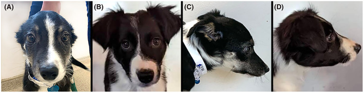
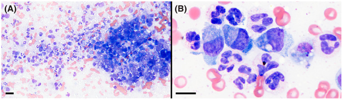
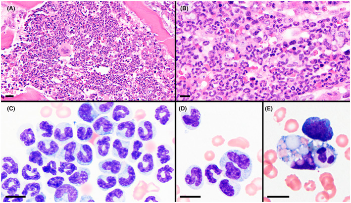
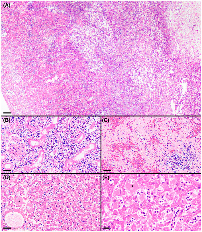
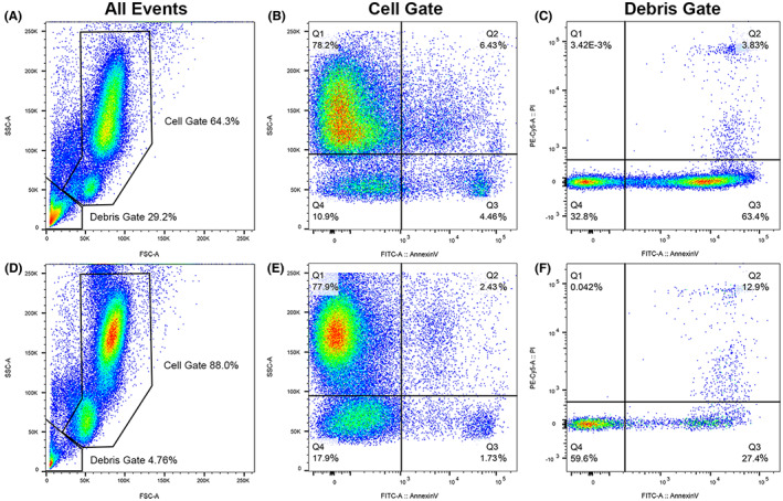
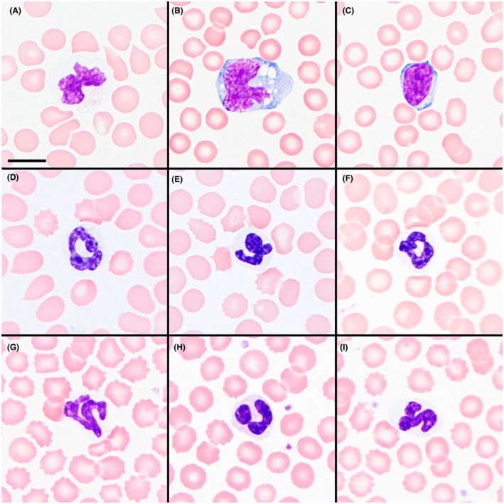
Trapped neutrophil syndrome is a rare congenital disease recognized in Border Collies and is characterized by persistent neutropenia with myeloid hyperplasia. The mechanism of neutropenia has not been described. We document the case of a young Border Collie diagnosed with trapped neutrophil syndrome based on clinical features, blood and bone marrow evaluation, and presence of the associated homozygous mutation. Results from flow cytometric and storage studies suggested lower neutrophil survival time. The dog had substantial neutrophilic inflammation in multiple organs, indicating that neutrophils could leave the marrow and enter tissues, making the term "trapped" neutrophil syndrome a misnomer.

Keywords: Cohen syndrome; VPS13B; bone marrow; ineffective myelopoiesis; neutropenia.


Conflict of interest statement

Authors declare no conflict of interest.

Figures

FIGURE 1
FIGURE 1
Abnormally narrow and elongated facial features in a 3‐month‐old Border Collie with trapped neutrophil syndrome (A and C) compared to a clinically healthy age‐matched dog of the same breed (B and D)
FIGURE 2
FIGURE 2
Neutrophilic and macrophagic inflammation in a cytologic specimen of the liver of the affected dog. Wright stain, magnification bars = 20 μm (A), 10 μm (B)
FIGURE 3
FIGURE 3
Bone marrow core biopsy (A and B) and aspirate (C‐E) specimens showing high cellularity and myeloid hyperplasia. Maturation of the myeloid lineage was complete through segmented neutrophils with prominent band neutrophils in many regions (B and C). Few giant neutrophils (D) and rare neutrophil phagocytosis (E) were present. H&E stain of B5‐fixed tissue (A and B) and Wright stain (C‐E); magnification bars = 20 μm (A), 10 μm (B‐E)
FIGURE 4
FIGURE 4
Histologic specimens of ileum (A), renal cortex (B), spleen (C), and liver (D and E). Effacing transmural necrotizing and suppurative lesion within the ileum, with the serosa to the left and the ulcerated mucosa to the right (A). Marked suppurative inflammation within the renal cortex and associated tubular degeneration (B). Marked amyloid deposition throughout this region of the spleen (C). Moderate hepatic amyloid deposition (asterisks denote representative accumulations) and many sinusoidal leukocytes, mostly neutrophils, despite a low‐normal blood neutrophil concentration (D and E). H&E stain; magnification bars = 100 μm (A), 50 μm (B‐D), 20 μm (E)
FIGURE 5
FIGURE 5
Flow cytometric scattergrams of blood leukocytes from the TNS dog (A‐C) and 1 representative control dog (D‐F). Initial gating (A and D) included 100 000 events for each dog and was based on forward scatter (horizontal axes), a representation of cell size, and side scatter (vertical axes), a representation of cell complexity into cell and debris gates; the affected dog had increased debris. The cell gate (B and E) shows expression of annexin V, a marker of cell death, on the horizontal axes, and side scatter on the vertical axes. The affected dog had an increased percentage of cells expressing annexin V compared to control dogs (upper and lower right quadrants). The top and bottom cell clouds in A, B, D, and E represent neutrophils and lymphocytes, respectively; monocytes appear as a subpopulation overlapping the bottom of the neutrophil cloud (labeling for each cell type not shown). The debris gate (C and F) shows expression of annexin V on the horizontal axes and propidium iodide, an additional marker of cell death, on the vertical axes. The affected dog had a greater percentage of annexin Vpos/propidium iodideneg events (lower right quadrant) compared to each of 4 control dogs
FIGURE 6
FIGURE 6
EDTA‐anticoagulated whole blood after 12 hours of refrigeration depicting a representative neutrophil (A), monocyte (B), and lymphocyte (C) from the affected dog compared to segmented neutrophils (D‐I) from 6 control dogs after the same storage time. Wright stain; magnification bar = 10 μm

References

    1. Allan FJ, Thompson KG, Jones BR, Burbidge HM, McKinley R. Neutropenia with a probable hereditary basis in border collies. N Z Vet J. 1996;44(2):67‐72. - PubMed
    1. Hegler AK, Grooters AM, Dehghanpir SD, Gallaher RA, Gaschen LE. Trapped neutrophil syndrome in a border collie. J Am Anim Hosp Assoc. 2020;56(3):e563‐04. - PubMed
    1. Gans Z. Confirmed trapped neutrophil syndrome in a border collie puppy in Israel. Isr J Vet Med. 2015;70(2):45‐48.
    1. Mason SL, Jepson R, Maltman M, Batchelor DJ. Presentation and management of trapped neutrophil syndrome (TNS) in UK border collies. J Small Anim Pract. 2014;55(1):57‐60. - PubMed
    1. Mizukami K, Shoubudani T, Nishimoto S, et al. Trapped neutrophil syndrome in a border collie dog: clinical, clinico‐pathologic, and molecular findings. J Vet Med Sci. 2012;74(6):797‐800. - PubMed

Publication types

Grants and funding