. 2022 Oct 1;149(19):dev200683.
doi: 10.1242/dev.200683. Epub 2022 Oct 13.

Loss of function of the maternal membrane oestrogen receptor ERα alters expansion of trophoblast cells and impacts mouse fertility

Affiliations

Loss of function of the maternal membrane oestrogen receptor ERα alters expansion of trophoblast cells and impacts mouse fertility

Mariam Rusidzé et al. Development. .

Abstract

The binding of 17β-oestradiol to oestrogen receptor alpha (ERα) plays a crucial role in the control of reproduction, acting through both nuclear and membrane-initiated signalling. To study the physiological role of membrane ERα in the reproductive system, we used the C451A-ERα mouse model with selective loss of function of membrane ERα. Despite C451A-ERα mice being described as sterile, daily weighing and ultrasound imaging revealed that homozygous females do become pregnant, allowing the investigation of the role of ERα during pregnancy for the first time. All neonatal deaths of the mutant offspring mice resulted from delayed parturition associated with failure in pre-term progesterone withdrawal. Moreover, pregnant C451A-ERα females exhibited partial intrauterine embryo arrest at about E9.5. The observed embryonic lethality resulted from altered expansion of Tpbpa-positive spiral artery-associated trophoblast giant cells into the utero-placental unit, which is associated with an imbalance in expression of angiogenic factors. Together, these processes control the trophoblast-mediated spiral arterial remodelling. Hence, loss of membrane ERα within maternal tissues clearly alters the activity of invasive trophoblast cells during placentogenesis. This previously unreported function of membrane ERα could open new avenues towards a better understanding of human pregnancy-associated pathologies.

Keywords: Fertility; Membrane signalling; Oestrogen receptor ERα (ESR1); Parturition; Spiral arterial remodelling; Trophoblast cells.


Conflict of interest statement

Competing interests The authors declare no competing or financial interests.

Figures

Fig. 1.
Fig. 1.
C451A-ERα females can become pregnant but exhibit parturition failure that results in absence of offspring. (A,B) Evolution of body weight gain from embryonic day (E) 0.5 to E14.5 (A) and E14.5 to E27.5 (B) in WT-ERα and C451A-ERα females, mated with WT-ERα males. Two-way ANOVA with Tukey's post-hoc test. aP<0.05 for WT-ERα versus not pregnant; bP<0.05 for C451A-ERα versus not pregnant; cP<0.05 for WT-ERα versus C451A-ERα; dP<0.05 for C451A-ERα versus C451A-ERα with low weight gain; eP<0.05 for WT-ERα versus C451A-ERα with low weight gain; fP<0.05 for C451A-ERα with low weight gain versus not pregnant. Mice were considered to be pregnant if they had gained more than 1 g by E7 or as non-pregnant if they had gained less than 1 g by E7. However, if they had gained more than 1 g by E7 but their weight gain had not doubled by E14, mice were classified as low weight gain. Resorptions were detected in the uteri of two mice sacrificed at E14, demonstrating that they could not be considered to be pregnant nor non-pregnant. (C) Gestational lengths of pregnant WT-ERα and C451A-ERα females that gave birth between E18.5 and E26.5. ****P<0.0001 (Mann–Whitney test). (D) Number of total (left) and live (middle) pups per litter found in nests on postnatal day 1 and percentage of pregnant females (right) after three consecutive mating tests in WT-ERα (n=10) and C451A-ERα mice (n=12), mated with WT-ERα male for 7 days. Mann–Whitney tests or Fisher's exact test: **P<0.01 and ***P<0.001. Data are representative of three independent experiments. (E,F) Placental (E) and offspring body (F) weights of foetuses from C451A-ERα mice (n=8) and WT-ERα littermates (n=8) at E18.5 after caesarean sections. **P<0.01 (Mann–Whitney test). Each point corresponds to the means of all concepti per mother. (G) Circulating levels of 17β-oestradiol (E2) and progesterone in pregnant WT-ERα (n=8) and C451A-ERα (n=8) mice at E18.5. ***P<0.001 (Mann–Whitney test).
Fig. 2.
Fig. 2.
ERα-C451A mice exhibit intra-uterine embryonic lethality. (A-C) Number of total (A), and total and live (B,C) embryos per mother at E9.5 (A), E14.5 (B) and E18.5 (C), evidenced by ultrasound imaging. *P<0.05, **P<0.01 and ***P<0.001 (Mann–Whitney tests). Dots on the graphs represent numbers from a single mother.
Fig. 3.
Fig. 3.
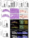
C451A-ERα pregnant females show placentomegaly due to dilated vascular channels in the junctional zone. (A) Percentages of aborted embryos observed in pregnant C451A-ERα (n=6) and WT-ERα (n=12) mice. *P<0.05, Mann–Whitney test. (B) Placenta thickness, diameter and weight at E14.5. *P<0.05, ***P<0.001, Mann–Whitney test. (C,D) Quantifications of junctional zone (C) and labyrinth (D) areas (mm2). Data include 32 placentas from 14 mothers (n for WT-ERα=6 and n for C451A-ERα=10). Dots represent the mean areas of all placentas obtained from the same mother. *P<0.05, Mann–Whitney test. (E) Transverse histological sections of whole placentas (left) collected from WT-ERα and C451A-ERα mice at E14.5 showing all major placental structural components. Magnified views of the junctional zone (Jz), delineated by a dotted line, are shown in the right inset. Db, decidua basalis; Jz, junctional zone; Lab, labyrinth. Scale bars: 2.5 mm (right) and 1 mm (left). (F,G) Respective quantification (G) with schematic illustration (F) of the surface of vascular channels (red area) in the junctional zone (dotted line) represented by average measurements per mother. The data highlight the dilated vascular tree of Jz in placentas of C451A-ERα mice (right, n=10), compared with the WT-ERα mice (left, n=6). ***P<0.001 Mann–Whitney test. (H) Double staining of MCT1 (green) and MCT4 (red) or MCT1 (green) and CD31 (red) mouse placental sections at E14.5. Labelling of maternal vasculature (CD31) and of apical (MCT1) and basal (MCT4) plasma membranes of labyrinthine syncytiotrophoblast cells. Scale bars: 2 mm (left); 100 μm (right). (I) Oxygen saturation values (SO2) of whole placental tissue at E14.5, represented by average measurements per mother. SO2 was measured in two to five live embryos per mother. WT-ERα (n=5) and C451A-ERα (n=5). Scale bars: 3 mm. (J) Examples of B-mode and photoacoustic-mode (PA) in vivo images of SO2 measurements at E14.5. The merge panel illustrates the custom colour map of oxygen saturation superimposed on the ultrasound image, where red corresponds to completely oxygenated blood and blue to completely deoxygenated blood. Scale bar: 2 mm.
Fig. 4.
Fig. 4.
Physiological remodelling of resistance uterine arteries is not affected by membrane ERα loss of function. (A) Organization of the uterine circulation in the pregnant mouse. Mouse pregnancy is supported by two uterine arteries, one on each side of the uterine horn (left and right). This arterial loop is generated from direct anastomoses of the uterine branch of the ovarian artery (OA) (cranially) and the uterine artery (UA) (caudally), resulting in counter bi-directional blood flow. The uterine artery further branches out to the arcuate artery (AA), the radial artery (RA) and the spiral artery (SA), which, respectively, supply the endometrium, decidua and placenta during pregnancy. Dotted rectangle outlines the uterine arterial segment that was used for this experiment. (B,C) Diameter (µm) of two fragments (UA and OA) of uterine arteries isolated from non-pregnant and pregnant WT-ERα and C451A-ERα mice at E9.5 after a stepwise increase in intraluminal pressure. Data show a significant pressure effect in pregnant and non-pregnant females in both genotypes. aP<0.0001 versus non-pregnant for wild-type mice; bP<0.01, cP<0.001 or dP<0.0001 versus non-pregnant for C451A-ERα mice following significant interaction in a two-way repeated measure ANOVA. Data are mean±s.e.m.
Fig. 5.
Fig. 5.
C451A-ERα mutation in maternal tissues results in dysregulation of genes associated with spiral arterial remodelling in mice. Real time RT-qPCR for Tpbpa, Prl7b1, Prl4a1, Prlc2c, Prl3d1, Pcdh12, Esr1, Pgr, Plgf, sFlt1/Vegfr-1, Vegf-a, Flt1/Vegfr-1, KDR/Vegfr-2, Ang2 and Egfl7 genes in whole implantation sites from WT-ERα and C451A-ERα mice (isolated at E9.5). Data include 59 samples, collected from seven WT-ERα and six C451A-ERα mice. Each violin plot represents a distribution of data corresponding to three to five samples obtained from the same mother. Nested t-test: *P<0.05, **P<0.01, ***P<0.001. Data are mean±s.e.m.
Fig. 6.
Fig. 6.
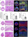
Histological analysis of concepti at E9.5 of WT-ERα and C451A-ERα pregnant females. (A-C) Histological images of middle sections of whole concepti at E9.5, including magnification on the left (A), and respective analyses of the placental surface (mm2) (B) and of the thickness of utero-placental unit (C) in C451A-ERα mice (n=5) compared with WT-ERα control (n=5). Dots represent the mean areas of three or four placentas per mother. SA, spiral artery; TGC, trophoblast giant cell; SpT, spongiotrophoblast; Lab, labyrinth; Ch, chorion. Yellow double-headed arrow indicates the thickness of the utero-placental unit. Scale bars: 2.5 mm, 500 µm and 250 µm (lowest to highest magnifications). *P<0.05, Mann–Whitney test. (D,E) Representative images of the trophoblast progenitor specific marker Tpbpa (green) and CK8 (red) immunostaining in serial sections of whole concepti at E9.5 of WT-ERα and C451A-ERα females. Lower and higher magnifications of the area outlined on the left are shown on the right with respective quantification of the cellular surface (mm2) positively immunoreactive for anti-Tpbpa antibody (E). Scale bars: 1 mm for images of concepti, 250 µm and 100 µm (higher and lower magnification, respectively). *P<0.05, Mann–Whitney test. (F) Representative images of Haematoxylin and Eosin staining of middle sections from some concepti of C451A-ERα females at E9.5 that display important disorganization of utero-placental unit showing growth arrest, accompanied by vascular dilatation, hyperaemia, oedema and even total embryo necrosis (asterisks). Scale bars: 2.5 mm. (G) Representative images of the trophoblast progenitor-specific marker Tpbpa (green) and CK8 (red) immunostaining in serial sections of concepti at E9.5 of C451A-ERα females where immunostaining of Tpbpa is totally absent. Scale bars: 1 mm (images of concepti); 250 μm (higher magnification images).

References

    1. Adamson, S. L., Lu, Y., Whiteley, K. J., Holmyard, D., Hemberger, M., Pfarrer, C. and Cross, J. C. (2002). Interactions between trophoblast cells and the maternal and fetal circulation in the mouse placenta. Dev. Biol. 250, 358-373. 10.1006/dbio.2002.0773 - DOI - PubMed
    1. Adlanmerini, M., Solinhac, R., Abot, A., Fabre, A., Raymond-Letron, I., Guihot, A.-L., Boudou, F., Sautier, L., Vessières, E., Kim, S. H.et al. (2014). Mutation of the palmitoylation site of estrogen receptor α in vivo reveals tissue-specific roles for membrane versus nuclear actions. Proc. Natl. Acad. Sci. USA 111, E283-E290. 10.1073/pnas.1322057111 - DOI - PMC - PubMed
    1. Adlanmerini, M., Fébrissy, C., Zahreddine, R., Vessières, E., Buscato, M., Solinhac, R., Favre, J., Anquetil, T., Guihot, A.-L., Boudou, F.et al. (2020). Mutation of Arginine 264 on ERα (Estrogen Receptor Alpha) selectively abrogates the rapid signaling of estradiol in the endothelium without altering fertility. Arterioscler. Thromb. Vasc. Biol. 40, 2143-2158. 10.1161/ATVBAHA.120.314159 - DOI - PubMed
    1. Albrecht, E. D. and Pepe, G. J. (2010). Estrogen regulation of placental angiogenesis and fetal ovarian development during primate pregnancy. Int. J. Dev. Biol. 54, 397-408. 10.1387/ijdb.082758ea - DOI - PMC - PubMed
    1. Albrecht, E. D., Robb, V. A. and Pepe, G. J. (2004). Regulation of placental vascular endothelial growth/permeability factor expression and angiogenesis by estrogen during early baboon pregnancy. J. Clin. Endocrinol. Metab. 89, 5803-5809. 10.1210/jc.2004-0479 - DOI - PubMed

Publication types