. 2022 Oct;8(10):mgen000869.
doi: 10.1099/mgen.0.000869.

Trichoderma atroviride hyphal regeneration and conidiation depend on cell-signaling processes regulated by a microRNA-like RNA

Affiliations

Trichoderma atroviride hyphal regeneration and conidiation depend on cell-signaling processes regulated by a microRNA-like RNA

José M Villalobos-Escobedo et al. Microb Genom. 2022 Oct.

Abstract

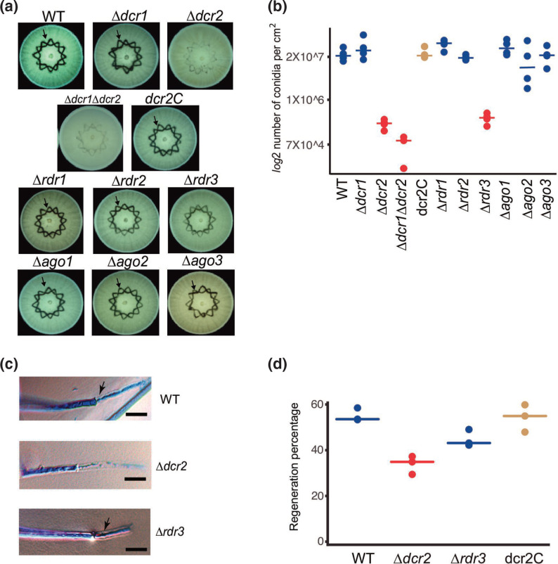
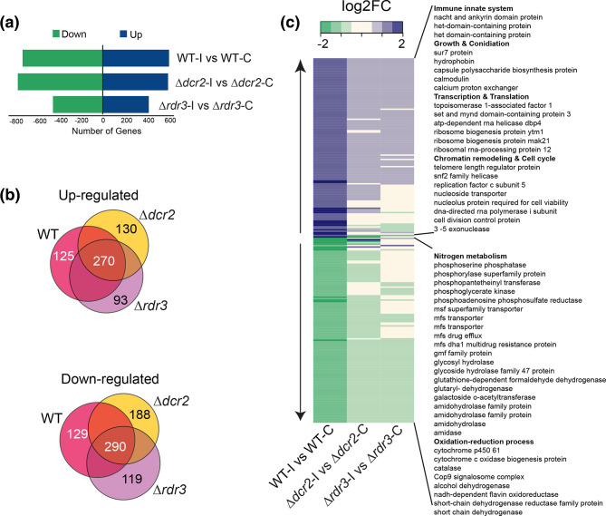
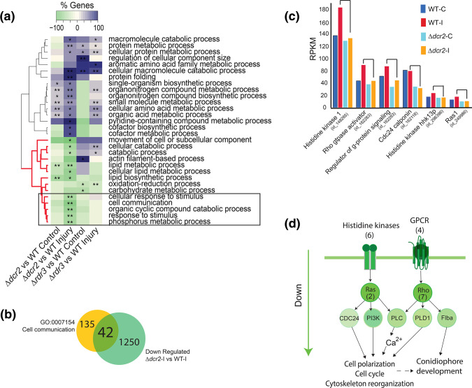
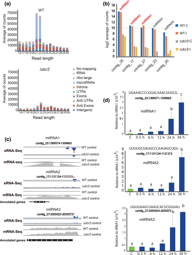
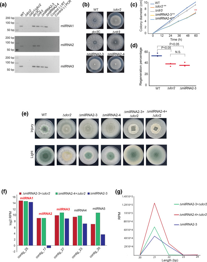
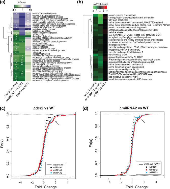
The ability to respond to injury is essential for the survival of an organism and involves analogous mechanisms in animals and plants. Such mechanisms integrate coordinated genetic and metabolic reprogramming events requiring regulation by small RNAs for adequate healing of the wounded area. We have previously reported that the response to injury of the filamentous fungus Trichoderma atroviride involves molecular mechanisms closely resembling those of plants and animals that lead to the formation of new hyphae (regeneration) and the development of asexual reproduction structures (conidiophores). However, the involvement of microRNAs in this process has not been investigated in fungi. In this work, we explore the participation of microRNA-like RNAs (milRNAs) molecules by sequencing messenger and small RNAs during the injury response of the WT strain and RNAi mutants. We found that Dcr2 appears to play an important role in hyphal regeneration and is required to produce the majority of sRNAs in T. atroviride. We also determined that the three main milRNAs produced via Dcr2 are induced during the damage-triggered developmental process. Importantly, elimination of a single milRNA phenocopied the main defects observed in the dcr2 mutant. Our results demonstrate the essential role of milRNAs in hyphal regeneration and asexual development by post-transcriptionally regulating cellular signalling processes involving phosphorylation events. These observations allow us to conclude that fungi, like plants and animals, in response to damage activate fine-tuning regulatory mechanisms.

Keywords: RNA-seq; RNAi machinery; conidiation; filamentous fungi; hyphal regeneration; milRNAs; signaling; small RNAs; transcriptome.


Conflict of interest statement

The authors declare that they have no conflicts of interests.

Figures

Fig. 1.
Fig. 1.
The response to mechanical damage is affected in RNAi mutants. (a) Conidiation response to injury of the WT strain and RNAi mutants. Colonies of the indicated strains growing on PDA were damaged with a cookie mould and photographed 36 h later. The arrow indicates the areas of accumulation of conidia in response to injury (dark). (b) The graph shows the yield of conidia in response to injury of four biological replicates. (c) Hyphal regeneration 1 h after injury. Hyphae were stained with lactophenol cotton blue and examined by light microscopy. Arrows point to the new hyphae. Scale bar=10 µM. (d) Percentage of hyphae that regenerate after injury for the indicated strains. Three biological replicates were used, counting 50 hyphae per strain. In (b) and (d), a one-way ANOVA test, followed by Tukey honest significant differences was applied. Highlights in red indicate that there was a statistically significant difference when comparing the indicated strains with the WT (P<0.05).
Fig. 2.
Fig. 2.
Genes involved in nitrogen metabolism and redox balance are regulated by RNAi. (a) Number of differentially expressed genes in the WT, Δdcr2 and Δrdr3 subjected to injury, compared to their corresponding undamaged control (FDR <0.05, fold-change >1). (b) Venn diagrams of the overexpressed and repressed genes in the different comparisons (blue=WT, yellow = Δdcr2 and green = Δrdr3), indicating the number of exclusive genes for each strain and those shared by all strains. (c) Heat map of the expression profile of the 254 genes differentially expressed only in the WT strain (125 up-regulated and 129 down-regulated genes), and their behavior in the Δdcr2 and Δrdr3 strains.
Fig. 3.
Fig. 3.
dcr2 regulates signaling processes independently of rdr3. (a) Clustering of GO terms-enrichment analysis belonging to Biological Process (FDR <0.01 **; FDR <0.05 *) in a paired comparison of the Δdcr2 and Δdcr3 mutants vs the WT strain in the control and injury conditions. (b) Overlap between the genes contained in the cell communication GO-term and the genes repressed specifically in the Δdcr2 in response to injury compared to the WT strain. (c) Expression profile of genes involved in signaling in the WT strain and the Δdcr2 mutant, both in the control condition and in response to injury. (d) Model of signaling pathways repressed at the transcriptional level in the dcr2 mutant.
Fig. 4.
Fig. 4.
Biogenesis of milRNAs depends on dicer-2 in T. atroviride. (a) Genomic annotation and size distribution of the sRNA reads, read counts are the average across three replicates of the control condition. (b) Expression profile 30 min after injury of the five predicted milRNAs counted using ShortStack. The y-axis scale shows the average of reads in logarithm base 2 comparing WT vs Δdcr2 (FDR <0.05). (c) Visualization of the genomic loci for milRNAs 1, 2 and 3. In blue, milRNAs (sRNA-Seq) and in grey likely pri-milRNA transcription (mRNA-Seq). Red stars indicate the regions of the mature milRNAs that were used for the stem-loop RT-qPCR. The black bars at the bottom represent potential coding regions and the white arrows show the direction of transcription (annotated genes). (d) Time course of the expression of milRNA1, milRNA2 and milRNA3 during injury-induced conidiation by stem-loop RT-qPCR, green bars show control expression without injury, blue bars show expression after injury (sequences of the milRNAs are shown at the top). The expression level is expressed relative to that of tRNA1, which does not change under these conditions. Three biological and five technical replicates were performed. Error bars represent ±sem. A one-way ANOVA test, followed by Tukey honest significant differences was used, different letters indicate significant differences (P<0.05).
Fig. 5.
Fig. 5.
Mutation of milRNA2 blocks asexual development in response to injury. (a) Gel electrophoresis analysis of stem-loop RT-PCR products of the three most abundant milRNAs in the indicated RNAi mutants and two independent milRNA2 mutants. (b) Photographs show colonies of the indicated strains after 72 h of growth. (c) Time course of colony growth of the indicated strains. (b) and (c) Colonies were grown on PDA in darkness. Error bars represent ±sem. A one-way ANOVA test, followed by Tukey honest significant differences was used (**P <0.01). (d) The graph shows the percentage of hyphae that regenerate after injury for the indicated strains. 50 hyphae in total for each replicate were examined and three biological replicates (points) were performed per strain, lines indicate the average regeneration percentage. A one-way ANOVA test, followed by Tukey honest significant differences was used. Highlights in red indicate that there is a statistically significant difference when comparing the indicated strain with the WT (P <0.05). (e) Photographs show the conidiation phenotypes in response to injury of the WT strain, Δdcr2, ΔmilRNA2, and the dikaryon resulting from the fusion of Δdcr2/ΔmilRNAs. The experiment was performed on PDA and in complete darkness. (f) Expression level (reads per million) of the five milRNAs predicted in both the mutant and the dikaryons of milRNA2. (g) Length distribution of sRNA reads between 20 and 24 nt in the milRNA2 mutant, as well as the two dykarions.
Fig. 6.
Fig. 6.
Transcriptome analysis of milRNA2 mutant in response to injury and microRNA-target prediction. (a) Clustering of GO-enrichment analysis belonging to biological process (FDR <0.01 **, FDR <0.05 *) in the general comparison WT vs Δdcr2, ΔmilRNA2 and Δrdr3 mutants. (b) Heatmap of the expression profile of the genes affected by the mutation of milRNA2 and dcr2 (FDR <0.05). (c) Cumulative frequency curve of the expression profile of target genes of the three main milRNAs in the comparison Δdcr2 vs WT, the x-axis shows the log2-Fold-change, the black curve represents the expression profile of all the genes. (d) Cumulative frequency curve of the expression profile of target genes of the three main milRNAs in the comparison ΔmilRNA2 vs WT, the x-axis shows the log2-Fold-change, the black curve represents the expression profile of all the genes. The black arrow indicates the tendency of a group of milRNA2 target genes to be induced with respect to the average distribution of all the genes in the transcriptome.

Similar articles

Cited by

References

    1. Villalobos-Escobedo JM, Martínez-Hernández JP, Pelagio-Flores R, González-De la Rosa PM, Carreras-Villaseñor N, et al. 2022. Trichoderma atroviride hyphal regeneration and conidiation depend on cell-signaling processes regulated by a microRNA?like RNA. Figshare. - DOI - PMC - PubMed
    1. Ricci L, Srivastava M. Wound-induced cell proliferation during animal regeneration. Wiley Interdiscip Rev Dev Biol. 2018;7:e321. doi: 10.1002/wdev.321. - DOI - PubMed
    1. Birnbaum KD, Sánchez Alvarado A. Slicing across kingdoms: regeneration in plants and animals. Cell. 2008;132:697–710. doi: 10.1016/j.cell.2008.01.040. - DOI - PMC - PubMed
    1. Biggar KK, Storey KB. Insight into post-transcriptional gene regulation: stress-responsive microRNAs and their role in the environmental stress survival of tolerant animals. J Exp Biol. 2015;218:1281–1289. doi: 10.1242/jeb.104828. - DOI - PubMed
    1. Follert P, Cremer H, Béclin C. MicroRNAs in brain development and function: a matter of flexibility and stability. Front Mol Neurosci. 2014;7:5. doi: 10.3389/fnmol.2014.00005. - DOI - PMC - PubMed

Publication types

Supplementary concepts