

Evidence of D-shaped wounds in the intrasomatic bullet path: two case reports
- PMID: 36239911
- PMCID: PMC10014649
- DOI: 10.1007/s12024-022-00538-6
Evidence of D-shaped wounds in the intrasomatic bullet path: two case reports
Abstract
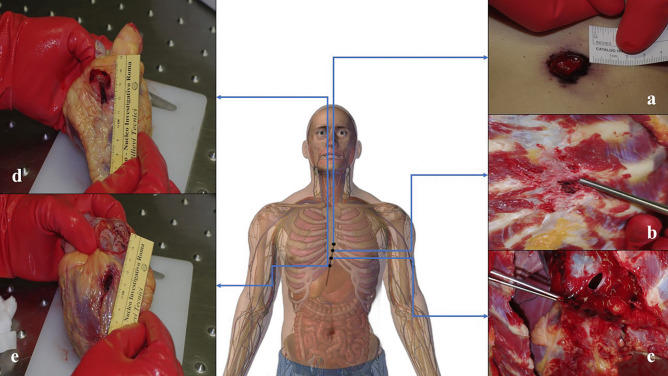
The final appearance of gunshot wounds is influenced by both the projectile's behavior from the muzzle to the terminal target and by the intrinsic characteristics of the anatomical compartments where the lesion(s) occur. The D-shaped morphology is an uncommon yet well-known finding in the forensic literature and has been described when the surface of impact with the skin is represented by the bullet's lateral projection. Two cases where D-shaped gunshot wounds were observed are hereby presented: in both cases, interaction with multiple intermediate targets (case 1) and a human intermediate target (case 2) had been documented and confirmed by the forensic examination. Despite the different dynamics of production, this peculiar morphology was described throughout most of the intrasomatic bullet path in both the victims. The discovery of D-shaped gunshot wounds can guide the forensic pathologist in the ballistic reconstruction of the event by supporting the hypothesis of an interaction with an intermediate target that has led to deviation from the initial trajectory and destabilization of the bullet associated with loss of kinetic energy.
Keywords: Atypical gunshot wounds; Bullet path; D-shaped gunshot wounds; Intermediated target; Ricochet.
© 2022. The Author(s).
Conflict of interest statement
The authors declare no competing interests.
Figures


References
-
- Humphrey C, Kumaratilake J. Ballistics and anatomical modelling–A review. Legal Med. 2016;23:21-9. 10.1016/j.legalmed.2016.09.002. - PubMed
-
- Madea B, Karger B. Handbook of forensic medicine. Chapter 20: Forensic Ballistics: Injuries from Gunshots, Explosives and Arrows, 1st Edition. Blackwell Pub. 2014.
-
- DiMaio VJ. Gunshot wounds: practical aspects of firearms, ballistics, and forensic techniques, Third Edition. CRC Press, 2015.
-
- Thali MJ, et al. Coins as intermediate targets: reconstructive analysis with synthetic body models. Am J Forensic Med Pathol. 2009;30(2):159–161. 10.1097/PAF.0b013e318187df63. - PubMed
-
- Karger B, Hoekstra A, Schmidt PF. Trajectory reconstruction from trace evidence on spent bullets. Int J Legal Med. 2001;115(1):16–22. 10.1007/s004140000202. - PubMed
Publication types
MeSH terms
LinkOut - more resources
Full Text Sources
