. 2022 Oct 3;5(10):e2236626.
doi: 10.1001/jamanetworkopen.2022.36626.

Classification of High-Grade Serous Ovarian Cancer Using Tumor Morphologic Characteristics

Affiliations

Classification of High-Grade Serous Ovarian Cancer Using Tumor Morphologic Characteristics

Katelyn F Handley et al. JAMA Netw Open. .

Abstract

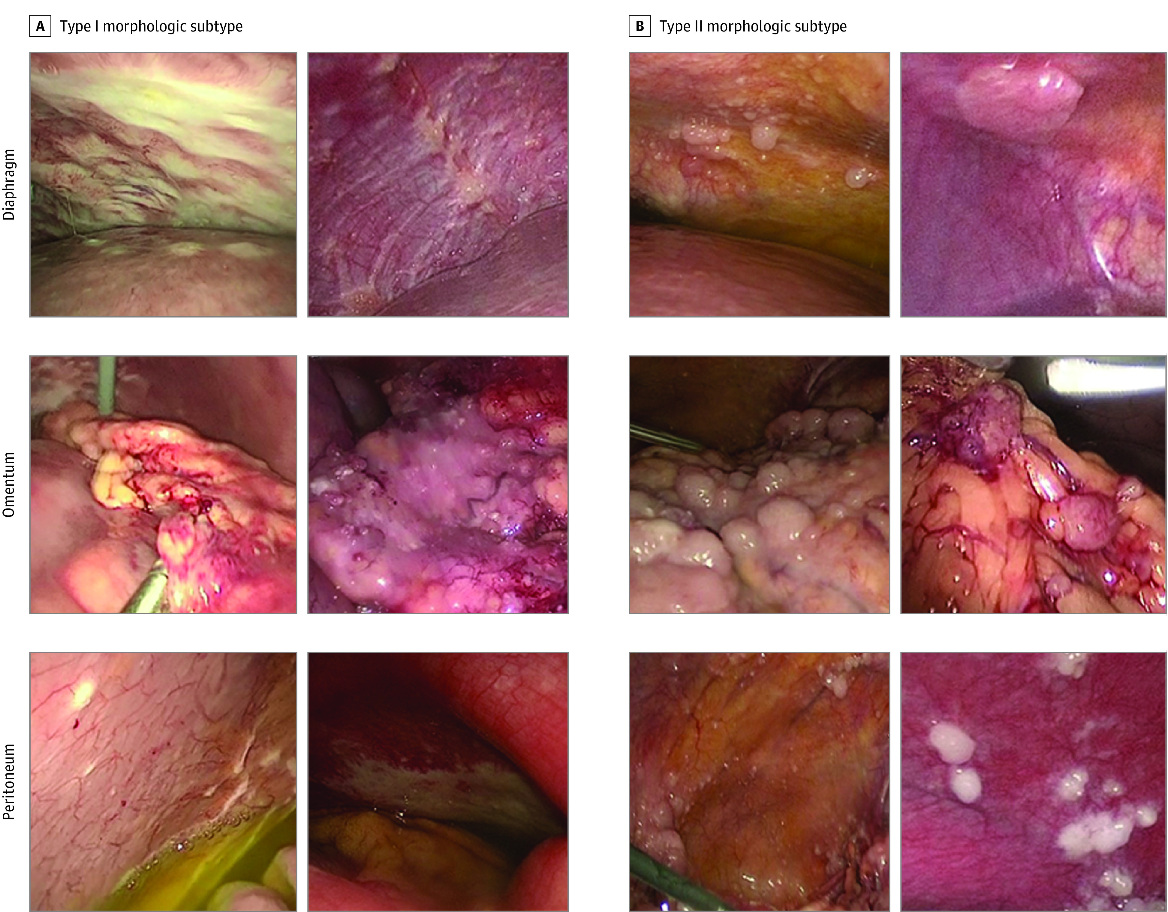
Importance: Despite similar histologic appearance among high-grade serous ovarian cancers (HGSOCs), clinical observations suggest vast differences in gross appearance. There is currently no systematic framework by which to classify HGSOCs according to their gross morphologic characteristics.

Objective: To develop and characterize a gross morphologic classification system for HGSOC.

Design, setting, and participants: This cohort study included patients with suspected advanced-stage ovarian cancer who presented between April 1, 2013, and August 5, 2016, to the University of Texas MD Anderson Cancer Center, a large referral center. Patients underwent laparoscopic assessment of disease burden before treatment and received a histopathologic diagnosis of HGSOC. Researchers assigning morphologic subtype and performing molecular analyses were blinded to clinical outcomes. Data analysis was performed between April 2020 and November 2021.

Exposures: Gross tumor morphologic characteristics.

Main outcomes and measures: Clinical outcomes and multiomic profiles of representative tumor samples of type I or type II morphologic subtypes were compared.

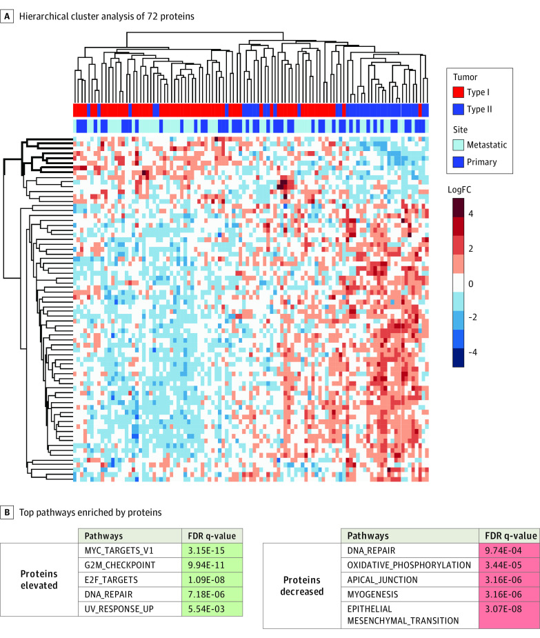
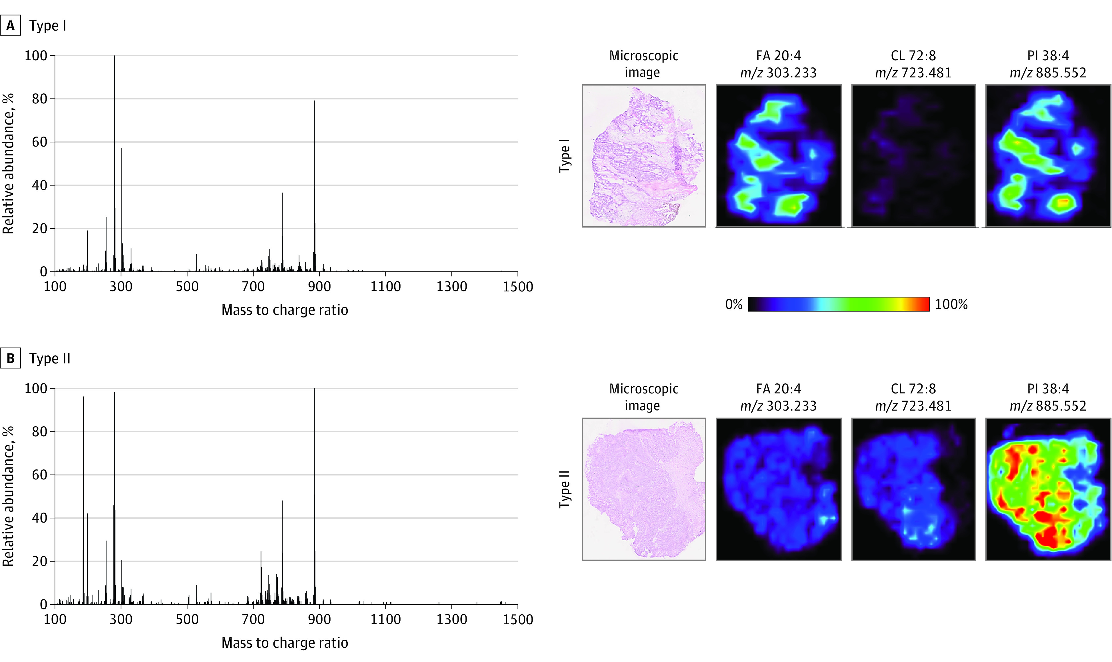
Results: Of 112 women (mean [SD] age 62.7 [9.7] years) included in the study, most patients (84% [94]) exhibited a predominant morphologic subtype and many (63% [71]) had a uniform morphologic subtype at all involved sites. Compared with those with uniform type I morphologic subtype, patients with uniform type II morphologic subtype were more likely to have a favorable Fagotti score (83% [19 of 23] vs 46% [22 of 48]; P = .004) and thus to be triaged to primary tumor reductive surgery. Similarly, patients with uniform type II morphologic subtype also had significantly higher mean (SD) estimated blood loss (639 [559; 95% CI, 391-887] mL vs 415 [527; 95% CI, 253-577] mL; P = .006) and longer mean (SD) operative time (408 [130; 95% CI, 350-466] minutes vs 333 [113; 95% CI, 298-367] minutes; P = .03) during tumor reductive surgery. Type I tumors had enrichment of epithelial-mesenchymal transition (false discovery rate [FDR] q-value, 3.10 × 10⁻²⁴), hypoxia (FDR q-value, 1.52 × 10⁻⁵), and angiogenesis pathways (FDR q-value, 2.11 × 10⁻²), whereas type II tumors had enrichment of pathways related to MYC signaling (FDR q-value, 2.04 × 10⁻⁹) and cell cycle progression (FDR q-value, 1.10 × 10⁻⁵) by integrated proteomic and transcriptomic analysis. Abundances of metabolites and lipids also differed between the 2 morphologic subtypes.

Conclusions and relevance: This study identified 2 novel, gross morphologic subtypes of HGSOC, each with unique clinical features and molecular signatures. The findings may have implications for triaging patients to surgery or chemotherapy, identifying outcomes, and developing tailored therapeutic strategies.


Conflict of interest statement

Conflict of Interest Disclosures: Dr Handley reported holding a pending provisional patent for Utilization of Immortalized B Cells to Identify SDCBP as a Novel Therapeutic Target in Ovarian Carcinoma. Mr Fellman reported receiving grants from the National Institutes of Health (NIH) during the conduct of the study. Dr Westin reported receiving grants from AstraZeneca, Bayer, Clovis Oncology, Mereo, Novartis, OncXerna, Bio-Path, Cotinga Pharmaceuticals, Roche/Genentech, and Zentalis, and receiving personal fees from AstraZeneca, Agenus, Clovis Oncology, Eisai, EQRX, ImmunoGen, Lilly, Mersana, Mereo, Roche/Genentech, Vincerx, Caris, and Zentalis outside the submitted work. Dr Maxwell reported receiving grants from the US Department of Defense during the conduct of the study. Dr Eberlin reported being a shareholder of MS Pen Technologies outside the submitted work and holding a patent for US20120295276A1, with royalties paid from Waters Corporation. Dr Fleming reported receiving personal fees from GSK and Immunogen outside the submitted work. Dr Sood reported receiving grants from the National Cancer Institute, Cancer Prevention and Research Institute of Texas (CPRIT), American Cancer Society (ACS), and Ovarian Cancer Research Alliance (OCRA) during the conduct of the study; receiving personal fees from AstraZeneca, GSK, Merck, Kiyatec, and Onxeo; being a shareholder with BioPath outside the submitted work; and holding a patent for EGFL6 antibody, licensed. No other disclosures were reported.

Figures

Figure 1.
Figure 1.. Representative Laparoscopic Images
Examples of type I and type II morphologic subtype of the diaphragm, omentum, and peritoneum.
Figure 2.
Figure 2.. Differential Protein Expression Analysis of High-Grade Serous Ovarian Cancer (HGSOC) Type I and Type II Tumors
A, Hierarchical cluster analysis of 72 proteins significantly altered between type II (n = 40) and type I (n = 63) HGSOC primary and metastatic tumors (linear models for microarray data [LIMMA]-adjusted P < .05). B, Top pathways enriched by proteins with significantly higher or lower expression in type II tumors than in type I tumors (LIMMA-adjusted P < .01). FC indicates fold change; FDR, false discovery rate.
Figure 3.
Figure 3.. Representative Desorption Electrospray Ionization Mass Spectra and Ion Images for Primary Tissues of Type I and Type II Morphologic Subtypes
In the ion images, red areas represent the highest relative abundances (100%), and black areas represent the lowest relative abundances (0%). Lipid species are described by their numbers of fatty acid chain carbons and double bonds. CL indicates cardiolipin; FA, fatty acid; PI, phosphatidylinositol.

References

    1. von Elm E, Altman DG, Egger M, Pocock SJ, Gøtzsche PC, Vandenbroucke JP; STROBE Initiative . The Strengthening the Reporting of Observational Studies in Epidemiology (STROBE) statement: guidelines for reporting observational studies. Ann Intern Med. 2007;147(8):573-577. doi:10.7326/0003-4819-147-8-200710160-00010 - DOI - PubMed
    1. Fleming ND, Nick AM, Coleman RL, et al. . Laparoscopic surgical algorithm to triage the timing of tumor reductive surgery in advanced ovarian cancer. Obstet Gynecol. 2018;132(3):545-554. doi:10.1097/AOG.0000000000002796 - DOI - PMC - PubMed
    1. Fagotti A, Ferrandina G, Fanfani F, et al. . A laparoscopy-based score to predict surgical outcome in patients with advanced ovarian carcinoma: a pilot study. Ann Surg Oncol. 2006;13(8):1156-1161. doi:10.1245/ASO.2006.08.021 - DOI - PubMed
    1. Fagotti A, Ferrandina G, Fanfani F, et al. . Prospective validation of a laparoscopic predictive model for optimal cytoreduction in advanced ovarian carcinoma. Am J Obstet Gynecol. 2008;199(6):642.e1-642.e6. doi:10.1016/j.ajog.2008.06.052 - DOI - PubMed
    1. Fagotti A, Vizzielli G, De Iaco P, et al. . A multicentric trial (Olympia-MITO 13) on the accuracy of laparoscopy to assess peritoneal spread in ovarian cancer. Am J Obstet Gynecol. 2013;209(5):462.e1-462.e11. doi:10.1016/j.ajog.2013.07.016 - DOI - PubMed

Publication types